Tuesday 27 May 2014

Migrating Stem Cells Possible New Focus for Stroke Treatment

Migrating Stem Cells Possible New Focus for Stroke Treatment
Tuesday, 27 May 2014

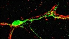
Brain pericyte. Credit: Lund University.
Two years ago, a new type of stem cell was discovered in the brain that has the capacity to form new cells. The same research group at Lund University in Sweden has now revealed that these stem cells, which are located in the outer blood vessel wall, appear to be involved in the brain reaction following a stroke.

The findings show that the cells, known as pericytes, drop out from the blood vessel, proliferate and migrate to the damaged brain area where they are converted into microglia cells, the brain's inflammatory cells.

Pericytes are known to contribute to tissue repair in a number of organs, and the researchers believe that their reparative properties could also apply to the brain. The study shows for the first time that pericytes are directly involved in the reaction of the brain tissue after stroke.

"Pericytes are a fascinating cell type with many different properties and found at high density in the brain. It was surprising that a pericyte subtype is so strongly activated after a stroke. The fact that pericytes can be converted into microglia, which have an important function in the brain after a stroke, was an unexpected finding that opens up a new possibility to influence inflammation associated with a stroke", said Gesine Paul-Visse, neurologist at Lund University and senior author of the study.

Using a green fluorescent protein bound to the pericytes, the researchers were able to track the cells' path to the damaged part of the brain. The migration takes place within a week after a stroke. When the cells reach the site of damage they are converted into microglia cells, the 'cleaners' of the central nervous system. Inflammation can, however, have both positive reparative effects and negative effects on the damaged tissue. The exact role of microglia cells in the regeneration after a stroke is not entirely clear, but we do know that pericytes play an important role in protecting the brain against disease and injury.

"We now need to elucidate how pericytes affect the brain's recovery following a stroke. Our findings put pericytes in focus as a new target for brain repair and future research will help us understand more about the brain's own defence and repair mechanisms."

There is an urgent need for new drugs that can alleviate the harmful effects of a stroke as current treatment possibilities using thrombolysis are limited to the first hours following a stroke.

"Because inflammation following a stroke is an event that continues after the acute stage, we hope that targeting pericytes in the sub-acute phase after stroke, i.e. within a longer time window following the onset of stroke, may influence the outcome", said Gesine Paul-Visse.

Source: Lund University 
Contact: Gesine Paul-Visse

Reference:
Brain pericytes acquire a microglial phenotype after stroke
Ilknur Özen, Tomas Deierborg, Kenichi Miharada, Thomas PAdel, Elisabet Englund, Guillem Genove and Gesine Paul 
Acta Neuropathologica. May 2014, 10.1007/s00401-014-1295-x
.........


For more on stem cells and cloning, go to CellNEWS at

Thursday 22 May 2014

New Insight into Stem Cell Development

New Insight into Stem Cell Development
Thursday, 22 May 2014

This is Professor Susanne Mandrup, University
of Southern Denmark. Credit: SDU.
The world has great expectations that stem cell research one day will revolutionize medicine. But in order to exploit the potential of stem cells, we need to understand how their development is regulated. Now researchers from University of Southern Denmark offer new insight.

Stem cells are cells that are able to develop into different specialized cell types with specific functions in the body. In adult humans these cells play an important role in tissue regeneration. The potential to act as repair cells can be exploited for disease control of e.g. Parkinson's or diabetes, which are diseases caused by the death of specialized cells. By manipulating the stem cells, they can be directed to develop into various specialized cell types. This however, requires knowledge of the processes that regulate their development.

Now Danish researchers from University of Southern Denmark report a new discovery that provides valuable insight into basic mechanisms of stem cell differentiation. The discovery could lead to new ways of making stem cells develop into exactly the type of cells that a physician may need for treating a disease.

"We have discovered that proteins called transcription factors work together in a new and complex way to reprogram the DNA strand when a stem cell develops into a specific cell type. Until now we thought that only a few transcription factors were responsible for this reprogramming, but that is not the case", explain postdoc Rasmus Siersbaek, Professor Susanne Mandrup and Ph.D. Atefeh Rabiee from Department of Biochemistry and Molecular Biology at the University of Southern Denmark.

"An incredibly complex and previously unknown interplay between transcription factors takes place at specific locations in the cell's DNA, which we call 'hotspots'. This interplay at 'hotspots' appears to be of great importance for the development of stem cells. In the future it will therefore be very important to explore these 'hotspots' and the interplay between transcription factors in these regions in order to better understand the mechanisms that control the development of stem cells", explains Rasmus Siersbaek.

"When we understand these mechanisms, we have much better tools to make a stem cell develop in the direction we wish", he says.

Siersbaek, Mandrup and their colleagues made the discovery while studying how stem cells develop into fat cells. The Mandrup research group is interested in this differentiation process, because fundamental understanding of this will allow researchers to manipulate fat cell formation.

"We know that there are two types of fat cells; brown and white. The white fat cells store fat, while brown fat cells actually increase combustion of fat. Brown fat cells are found in especially infants, but adults also have varying amounts of these cells.”

"If we manage to find ways to make stem cells develop into brown rather than white fat cells, it may be possible to reduce the development of obesity. Our findings open new possibilities to do this by focusing on the specific sites on the DNA where proteins work together", the researchers explain.

Details of the study
The researchers worked with a well-known cell line that can be induced to develop into fat cells in about a week in the laboratory using a specific cocktail of hormones. It has been known for many years that this process is regulated by proteins in the cells called transcription factors that control which genes are turned on and off during development of the fat cell.

So far it has been unclear how all these proteins work together to create a fat cell. In this paper, the researchers report that transcription factors bind together to special places in the cell's genome called ‘hotspots’. Here transcription factors 'talk' to each other and cooperate in controlling which genes are switched on and off. 'Hotspots' therefore act as key hubs in the genome where different signals are integrated on a small piece of DNA.

The researchers further showed that there are often several of these 'hotspots' close to each other and that they work together to form large so-called 'super-enhancers'. These 'super-enhancers' seem to be extra important in order to activate the right genes during fat cell development.

This study thus shows an extremely high degree of cooperativity between transcription factors at the level of DNA, which seems to be very important for directing the development of stem cells.

Contact: Birgitte Svennevig

References:
Molecular Architecture of Transcription Factor Hotspots in Early Adipogenesis 
Rasmus Siersbæk, Songjoon Baek, Atefeh Rabiee, Ronni Nielsen, Sofie Traynor, Nicholas Clark, Albin Sandelin, Ole N. Jensen, Myong-Hee Sung, Gordon L. Hager and Susanne Mandrup

Transcription Factor Cooperativity in Early Adipogenic Hotspots and Super-Enhancers
Rasmus Siersbæk, Atefeh Rabiee, Ronni Nielsen, Simone Sidoli, Sofie Traynor, Anne Loft, Lars La Cour Poulsen, Adelina Rogowska-Wrzesinska, Ole N. Jensen, and Susanne Mandrup
.........


For more on stem cells and cloning, go to CellNEWS at

A New Genetic Switching Element in Stem Cells

A New Genetic Switching Element in Stem Cells
Thursday, 22 May 2014

Slight modifications in their genome sequences play a crucial role in the conversion of pluripotent stem cells into various differentiated cell types. A team at Ludwig-Maximilians-Universitaet (LMU) in Munich has now identified the factor responsible for one class of modification.

Professor Thomas Carell from the Department
of Chemistry at LMU.
Every cell contains stored hereditary information, encoded in the sequence of nucleobases that make up its DNA. However, in any given cell type, only a fraction of this information is actually used. Which genes are activated and which are turned off is in part determined by a second tier of information which is superimposed on the nucleotide sequences that provide the blueprints for protein synthesis. This so-called epigenetic level of control is based on the localized, and in principle reversible, attachment of simple chemical tags to specific nucleotides in the genome. This system plays a major role in the regulation of gene activity, and enables the selective expression of different functions in differentiated cell types.

This explains why such DNA modifications play a major role in the differentiation of stem cells.

“Several unusual nucleobases have been found in the genomes of stem cells, which are produced by targeted chemical modification of the known building blocks of DNA. These ‘atypical’ bases are thought to be important in determining what types of differentiated cells can be derived from a given stem cell line,” says Professor Thomas Carell from the Department of Chemistry at LMU.

All of the unconventional bases so far discovered are derived from the same standard base – cytosine. Furthermore, Carell and his team have shown in earlier work that so-called Tet enzymes are always involved in their synthesis.

Base oxidation regulates gene activity
In cooperation with colleagues at LMU, as well as researchers based in Berlin, Basel and Utrecht, Carell and his group have now shown, for the first time, that a standard base other than cytosine is also modified in embryonic stem cells of mice. Moreover, Tet is at work here too.

“During the development of specialized tissues from stem cells, enzymes belonging to the Tet family also oxidize the thymidine base, as we have now shown with the aid of highly sensitive analytical methods based on mass spectrometry. The product of the reaction, hydroxymethyluracil, was previously – and as it now turns out, erroneously – thought to be synthesized by a different pathway,” Carell explains.

The precise function of hydroxymethyluracil remains unclear. However, using an innovative method for the identification of factors capable of binding to and “reading” the chemical tags that characterize unconventional DNA bases, Carell and colleagues have shown that stem cells contain specific proteins that recognize hydroxymethyluracil, and could therefore contribute to the regulation of gene activity in these cells.

“We hope that these new insights will make it possible to modulate the differentiation of stem cells – causing them to generate cells of a particular type,” says Carell.

“It would be wonderful if we were one day able to generate whole organs starting from differentiated cells produced, on demand, by stem cell populations.”

Contact: Luise Dirscherl

Reference:
Tet oxidizes thymine to 5-hydroxymethyluracil in mouse embryonic stem cell DNA
Toni Pfaffeneder, Fabio Spada, Mirko Wagner, Caterina Brandmayr, Silvia K Laube, David Eisen, Matthias Truss, Jessica Steinbacher, Benjamin Hackner, Olga Kotljarova, David Schuermann, Stylianos Michalakis, Olesea Kosmatchev, Stefan Schiesser, Barbara Steigenberger, Nada Raddaoui, Gengo Kashiwazaki, Udo Müller, Cornelia G Spruijt, Michiel Vermeulen, Heinrich Leonhardt, Primo Schär, Markus Müller & Thomas Carell
Nature Chemical Biology Published online 18 May 2014, doi:10.1038/nchembio.1532
.........


For more on stem cells and cloning, go to CellNEWS at

Wednesday 21 May 2014

Functional Nerve Cells from Skin Cells

Functional Nerve Cells from Skin Cells
Wednesday, 21 May 2014

A new method of generating mature nerve cells from skin cells could greatly enhance understanding of neurodegenerative diseases, and could accelerate the development of new drugs and stem cell-based regenerative medicine.

These are mature nerve cells generated from
human cells using enhanced transcription 
factors. Credit: Fahad Ali.
The nerve cells generated by this new method show the same functional characteristics as the mature cells found in the body, making them much better models for the study of age-related diseases such as Parkinson's and Alzheimer's, and for the testing of new drugs.

Eventually, the technique could also be used to generate mature nerve cells for transplantation into patients with a range of neurodegenerative diseases.

By studying how nerves form in developing tadpoles, researchers from the University of Cambridge were able to identify ways to speed up the cellular processes by which human nerve cells mature. The findings are reported in the May 27th edition of the journal Development.

Stem cells are our master cells, which can develop into almost any cell type within the body. Within a stem cell, there are mechanisms that tell it when to divide, and when to stop dividing and transform into another cell type, a process known as cell differentiation. Several years ago, researchers determined that a group of proteins known as transcription factors, which are found in many tissues throughout the body, regulate both mechanisms.

More recently, it was found that by adding these proteins to skin cells, they can be reprogrammed to form other cell types, including nerve cells. These cells are known as induced neurons, or iN cells. However, this method generates a low number of cells, and those that are produced are not fully functional, which is a requirement in order to be useful models of disease: for example, cortical neurons for stroke, or motor neurons for motor neuron disease.

In addition, for age-related diseases such as Parkinson's and Alzheimer's, both of which affect millions worldwide, mature nerve cells which show the same characteristics as those found in the body are crucial in order to enhance understanding of the disease and ultimately determine the best way to treat it.

"When you reprogram cells, you're essentially converting them from one form to another but often the cells you end up with look like they come from embryos rather than looking and acting like more mature adult cells," said Dr Anna Philpott of the Department of Oncology, who led the research.

"In order to increase our understanding of diseases like Alzheimer's, we need to be able to work with cells that look and behave like those you would see in older individuals who have developed the disease, so producing more 'adult' cells after reprogramming is really important."

By manipulating the signals which transcription factors send to the cells, Dr Philpott and her collaborators were able to promote cell differentiation and maturation, even in the presence of conflicting signals that were directing the cell to continue dividing.

When cells are dividing, transcription factors are modified by the addition of phosphate molecules, a process known as phosphorylation, but this can limit how well cells can convert to mature nerves. However, by engineering proteins which cannot be modified by phosphate and adding them to human cells, the researchers found they could produce nerve cells that were significantly more mature, and therefore more useful as models for disease such as Alzheimer's.

Additionally, very similar protein control mechanisms are at work to mature important cells in other tissues such as pancreatic islets, the cell type that fails to function effectively in type 2 diabetes. As well as making more mature nerves, Dr Philpott's lab is now using similar methods to improve the function of insulin-producing pancreas cells for future therapeutic applications.

"We've found that not only do you have to think about how you start the process of cell differentiation in stem cells, but you also have to think about what you need to do to make differentiation complete - we can learn a lot from how cells in developing embryos manage this," said Dr Philpott.

Contact: Sarah Collins
.........


For more on stem cells and cloning, go to CellNEWS at

Monday 19 May 2014

Illuminating Neuron Activity in 3-D

New technique allows scientists to monitor the entire nervous system of a small worm
Sunday, 18 May 2014

Researchers at MIT and the University of Vienna have created an imaging system that reveals neural activity throughout the brains of living animals. This technique, the first that can generate 3-D movies of entire brains at the millisecond timescale, could help scientists discover how neuronal networks process sensory information and generate behaviour.

Head region and the majority of the brain of a
zebrafish larvae, as recorded and reconstructed
using the light-field microscope. Credit: MIT.
The team used the new system to simultaneously image the activity of every neuron in the worm Caenorhabditis elegans, as well as the entire brain of a zebrafish larva, offering a more complete picture of nervous system activity than has been previously possible.

"Looking at the activity of just one neuron in the brain doesn't tell you how that information is being computed; for that, you need to know what upstream neurons are doing. And to understand what the activity of a given neuron means, you have to be able to see what downstream neurons are doing," says Ed Boyden, an associate professor of biological engineering and brain and cognitive sciences at MIT and one of the leaders of the research team.

"In short, if you want to understand how information is being integrated from sensation all the way to action, you have to see the entire brain."

The new approach, described May 18 in Nature Methods, could also help neuroscientists learn more about the biological basis of brain disorders.

"We don't really know, for any brain disorder, the exact set of cells involved," Boyden says.

"The ability to survey activity throughout a nervous system may help pinpoint the cells or networks that are involved with a brain disorder, leading to new ideas for therapies."

Boyden's team developed the brain-mapping method with researchers in the lab of Alipasha Vaziri of the University of Vienna and the Research Institute of Molecular Pathology in Vienna. The paper's lead authors are Young-Gyu Yoon, a graduate student at MIT, and Robert Prevedel, a postdoc at the University of Vienna.

High-speed 3-D imaging
Neurons encode information — sensory data, motor plans, emotional states, and thoughts — using electrical impulses called action potentials, which provoke calcium ions to stream into each cell as it fires. By engineering fluorescent proteins to glow when they bind calcium, scientists can visualize this electrical firing of neurons. However, until now there has been no way to image this neural activity over a large volume, in three dimensions, and at high speed.

Scanning the brain with a laser beam can produce 3-D images of neural activity, but it takes a long time to capture an image because each point must be scanned individually. The MIT team wanted to achieve similar 3-D imaging but accelerate the process so they could see neuronal firing, which takes only milliseconds, as it occurs.

The new method is based on a widely used technology known as light-field imaging, which creates 3-D images by measuring the angles of incoming rays of light. Ramesh Raskar, an associate professor of media arts and sciences at MIT and an author of this paper, has worked extensively on developing this type of 3-D imaging. Microscopes that perform light-field imaging have been developed previously by multiple groups. In the new paper, the MIT and Austrian researchers optimized the light-field microscope, and applied it, for the first time, to imaging neural activity.

With this kind of microscope, the light emitted by the sample being imaged is sent through an array of lenses that refracts the light in different directions. Each point of the sample generates about 400 different points of light, which can then be recombined using a computer algorithm to recreate the 3-D structure.

"If you have one light-emitting molecule in your sample, rather than just refocusing it into a single point on the camera the way regular microscopes do, these tiny lenses will project its light onto many points. From that, you can infer the three-dimensional position of where the molecule was," says Boyden, who is a member of MIT's Media Lab and McGovern Institute for Brain Research.

Prevedel built the microscope at the IMP in Vienna, and Yoon devised the computational strategies that reconstruct the 3-D images.

Neurons in action
The researchers used this technique to image neural activity in the worm C. elegans, the only organism for which the entire neural wiring diagram is known. This 1-millimeter worm has 302 neurons, each of which the researchers imaged as the worm performed natural behaviours, such as crawling. They also observed the neuronal response to sensory stimuli, such as smells.

The downside to light field microscopy, Boyden says, is that the resolution is not as good as that of techniques that slowly scan a sample. The current resolution is high enough to see activity of individual neurons, but the researchers are now working on improving it so the microscope could also be used to image parts of neurons, such as the long dendrites that branch out from neurons' main bodies. They also hope to speed up the computing process, which currently takes a few minutes to analyse one second of imaging data.

The researchers also plan to combine this technique with optogenetics, which enables neuronal firing to be controlled by shining light on cells engineered to express light-sensitive proteins. By stimulating a neuron with light and observing the results elsewhere in the brain, scientists could determine which neurons are participating in particular tasks.

Contact: Sarah McDonnell

Reference:
Simultaneous whole-animal 3D imaging of neuronal activity using light-field microscopy
Robert Prevedel, Young-Gyu Yoon, Maximilian Hoffmann, Nikita Pak, Gordon Wetzstein, Saul Kato, Tina Schrödel, Ramesh Raskar, Manuel Zimmer, Edward S Boyden & Alipasha Vaziri
Nature Methods (2014), doi:10.1038/nmeth.2964
.........


For more on stem cells and cloning, go to CellNEWS at
http://cellnews-blog.blogspot.com/
Wednesday 7 May 2014

Study Urges Caution in Stem Cell Clinical Trials for Heart Attack Patients

Study Urges Caution in Stem Cell Clinical Trials for Heart Attack Patients
Wednesday, 07 May 2014

A new study in Nature challenges research data that form the scientific basis of clinical trials in which heart attack patients are injected with stem cells to try and regenerate damaged heart tissue.

Researchers at Cincinnati Children's Hospital Medical Center and the Howard Hughes Medical Institute (HHMI), report May 7 that cardiac stem cells used in ongoing clinical trials – which express a protein marker called c-kit – do not regenerate contractile heart muscle cells at high enough rates to justify their use for treatment.

This image from the laboratory of Jeffery
Molkentin, PhD, principal investigator on the
heart stem cell study in Nature, shows
cardiomyocytes that are stained in a red cardiac
protein. The cells were taken from the
hearts of mice that were bred so that any cells in
their body from the c-kit lineage could be
tracked by the scientists. C-kit-generated cells are
shown in green/yellow, helping show that c-kit
cells do not produce cardiomyocytes in sufficient
quantities to use them for the regeneration
of contractile heart muscle.
Including collaboration from researchers at Cedars-Sinai Heart Institute in Los Angeles and the University of Minnesota's Lillehei Heart Institute, the study uncovers new evidence in what has become a contentious debate in the field of cardiac regeneration, according to Jeffery Molkentin, PhD, study principal investigator and a cardiovascular molecular biologist and HHMI investigator at the Cincinnati Children's Heart Institute.

"Our data suggest any potential benefit from injecting c-kit-positive cells into the hearts of patients is not because they generate new contractile cells called cardiomyocytes," Molkentin said.

"Caution is warranted in further clinical testing of this method until the mechanisms in play here are better defined or we are able to dramatically enhance the potential of these cells to generate cardiomyocytes."

Numerous heart attack patients have already been treated with c-kit-positive stem cells that are removed from healthy regions of a damaged heart then processed in a laboratory, Molkentin explained. After processing, the cells are then injected into these patients' hearts. The experimental treatment is based largely on preclinical studies in rats and mice suggesting that c-kit-positive stem cells completely regenerate myocardial cells and heart muscle. Thousands of patients have also previously undergone a similar procedure for their hearts but with bone marrow stem cells.

Molkentin and his colleagues report those previous preclinical studies in rodents do not reflect what really occurs within the heart after injury, where internal regenerative capacity is almost non-existent. Molkentin also said that combined data from multiple clinical trials testing this type of treatment show most patients experienced a roughly 3-5 percent improvement in heart ejection fraction – a measurement of how forcefully the heart pumps blood. Data in the current Nature study suggest this small benefit may come from the ability of c-kit-positive stem cells in heart to cause the growth of capillaries, which improves circulation within the organ, but not by generating new cardiomyocytes.

"What we show in our study is that c-kit-positive stem cells from the heart like to make endothelial cells that form capillaries. But in their natural environment in the heart, these c-kit positive cells do not like to make cardiomyocytes," Molkentin said.

"They will produce cardiomyocytes, but at rates so low – roughly one in every 3,000 cells – it becomes meaningless."

The c-kit protein is expressed on the surface of progenitor cells originally identified in bone marrow. These c-kit expressing cells can generate multiple different cell types that are destined to help form specific organ tissues or other parts of the body. Given its presence in bone marrow, c-kit cells are also involved in the production of different types of immune system cells.

Researchers in the current study worked with two lines of genetically bred mice to see how efficiently c-kit-positive cardiac progenitor cells would regenerate cardiomyocytes in the hearts of the animals. The authors measured heart cell regeneration rates during the animals' embryonic development, during aging and after myocardial infarction (heart attack).

The mice were bred so that a fluorescent protein marker was permanently added to any cell that expressed the Kit gene, regardless of what it then turned into. This allowed the scientists to track the specific types and volumes of any c-kit-positive cells being generated in the animals, including in their hearts.

Test results showed that c-kit-positive cells originating in the heart generated new cardiomyocytes at a percentage (from baseline) of 0.03 or less. The authors go on to report that the percentage of new cardiomyocyte generation actually falls to below 0.008 when considering a natural process called cellular fusion – which in this instance involves c-kit-positive cells from the bone marrow or circulating immune system cells fusing with cardiomyocytes in the heart.

As a follow up to the current study, Molkentin and his colleagues are currently testing genes and protein growth factors that may be able to boost the rate of new cardiomyocyte generation from c-kit-positive stem cells. Because the current study shows that endogenous c-kit cells at least have some limited ability to regenerate contractile heart cells, Molkentin said it may be possible to find a method to enhance this ability genetically so the cells can eventually be used in a truly therapeutically beneficial manner to make new contractile activity in the heart.

Contact: Nick Miller

Reference:
c-kit+ cells minimally contribute cardiomyocytes to the heart
Jop H. van Berlo, Onur Kanisicak, Marjorie Maillet, Ronald J. Vagnozzi, Jason Karch, Suh-Chin J. Lin, Ryan C. Middleton, Eduardo Marbán & Jeffery D. Molkentin
Nature Published online 07 May 2014, doi:10.1038/nature13309
.........


For more on stem cells and cloning, go to CellNEWS at
http://cellnews-blog.blogspot.com/

Researchers Identify How Heart Stem Cells Orchestrate Regeneration

Exosomes, tiny 'bubbles' secreted by specialized cardiac stem cells, appear to carry 'instructions' that spur heart cells to regenerate following heart attack
Wednesday, 07 May 2014

Investigators at the Cedars-Sinai Heart Institute – whose previous research showed that cardiac stem cell therapy reduces scarring and regenerates healthy tissue after a heart attack in humans – have identified components of those stem cells responsible for the beneficial effects.

In a series of laboratory and lab animal studies, Heart Institute researchers found that exosomes, tiny membrane-enclosed "bubbles" involved in cell-to-cell communication, convey messages that reduce cell death, promote growth of new heart muscle cells and encourage the development of healthy blood vessels.

"Exosomes were first described in the mid-1980s, but we only now are beginning to appreciate their potential as therapeutic agents. We have found that exosomes and the cargo they contain are crucial mediators of stem cell-based heart regeneration, and we believe this might lead to an even more refined therapy using the 'active ingredient' instead of the entire stem cell," said Eduardo Marbán, MD, PhD, director of the Cedars-Sinai Heart Institute and a pioneer in developing investigational cardiac stem cell treatments.

"The concept of exosome therapy is interesting because it could potentially shift our strategy from living-cell transplantation to the use of a non-living agent," he added.

"Stem cells must be carefully preserved to keep them alive and functioning until the time of transplant, and there are some risks involved in cell transplantation. In contrast, exosome therapy may be safer and simpler and based on a product with a longer shelf life."

In lab experiments, the researchers isolated exosomes from specialized human cardiac stem cells and found that exosomes alone had the same beneficial effects as stem cells. Exosomes also produced the same post-heart attack benefits in mice, decreasing scar size, increasing healthy heart tissue and reducing levels of chemicals that lead to inflammation. Even when exosomes were injected in mice after heart attack scars were well-established, and traditionally viewed as "irreversible," they brought about multiple structural and functional benefits.

Exosomes transport small pieces of genetic material, called microRNAs, which enable cells to communicate with neighbouring cells to change their behaviour. The researchers pinpointed one such microRNA – one that is especially plentiful in cardiac stem cell exosomes – as responsible for some of the benefits. It is likely, they believe, that this and other microRNAs in the exosomes work together to produce the regenerative effects.

"The exosomes appear to contain the signalling information needed to regenerate healthy heart tissue, they are naturally able to permeate cells, and they have a coating that protects their payloads from degradation as they shuttle from cell to cell," said Marbán, senior author of an article in the May 6, 2014 Stem Cell Reports.

"Injecting exosomes derived from specialized cardiac stem cells may be an attractive alternative to the transplantation of living cells."

Marbán and his clinical and research teams in 2009 performed the first procedure in which a heart attack patient's heart tissue was used to grow specialized stem cells that were injected back into the heart. In 2012, they reported results of a clinical trial that found significant reduction in the size of heart attack-caused scars in patients who underwent the experimental stem cell procedure, compared to others who did not.

They also published findings from an animal study showing that the effect of stem cell therapy following heart attack is indirect – the stem cells themselves do not survive long after being placed in the heart, but they cause enduring effects by stimulating the rapid growth of surviving heart tissue and attracting stem cells already in the heart, which mature into functional heart cells.

The new study sheds light on the underlying mechanisms, crediting stem cell exosomes and the communications cargo they carry for orchestrating regeneration to repair heart attack damage.

The process to grow cardiac-derived stem cells was developed earlier by Marbán when he was on the faculty of Johns Hopkins University. The university has filed for a patent on that intellectual property and has licensed it to Capricor Inc., a biotechnology company in which Marbán is a founder and equity holder. Cedars-Sinai has filed for a patent for the exosome discovery and has licensed it to Capricor. The company provided no funding for this study.

Contact: Sally Stewart

Reference:
Exosomes as Critical Agents of Cardiac Regeneration Triggered by Cell Therapy
Ahmed Gamal-Eldin Ibrahim, Ke Cheng, Eduardo Marbán
Stem Cell Reports, 6 May 2014, Volume 2, Issue 5, p606–619
.........


For more on stem cells and cloning, go to CellNEWS at
http://cellnews-blog.blogspot.com/

One Step Closer to Cell Reprogramming

One Step Closer to Cell Reprogramming
Wednesday, 07 May 2014

In 2012, John B. Gurdon and Shinya Yamanaka were awarded the Nobel Prize in medicine for discovering that adult cells can be reprogrammed into pluripotent ones (iPS); the cells obtained are capable of behaving in a similar way to embryonic stem cells, and hence have enormous potential for regenerative medicine.

Cells with activated Wnt can no longer be
reprogrammed (in green) are located on the
periphery; cells that can be reprogrammed are
aggregated and can be seen in the centre of the
image (in red). Credit: CRG.
However, although there are many research groups around the world studying this process, it is still not completely understood, it is not totally efficient, and it is not safe enough to be used as the basis for a new cell therapy.

Now, researchers at the Centre for Genomic Regulation (CRG) in Barcelona have taken a very important step towards understanding cell reprogramming and its efficiency: they have discovered the key role of the Wnt signalling pathway in transforming adult cells into iPS cells.

"Generally, transcription factors are used to try to increase or decrease the cell reprogramming process. We have discovered that we can increase the efficiency of the process by inhibiting the Wnt route", explains Francesco Aulicino, a PhD student in the Reprogramming and Regeneration group, led by Maria Pia Cosma and co-author of the study that has just been published in Stem Cell Reports.

The Wnt signalling pathway is a series of biochemical reactions that are produced in cells. In frogs or lizards, for example, these reactions are those that allow their extremities to regenerate if the animal suffers an injury. Although in general, humans and mammals have lost this regenerative capacity, the Wnt pathway is involved in numerous processes during embryonic development and cell fusion, as it is in reprogramming.

The researchers have studied how the Wnt route behaves throughout the entire process of transforming cells into iPS cells, which usually lasts two weeks. It is a very dynamic process that produces oscillations from the pathway, which is not active all the time.

"We have seen that there are two phases and that in each one of them, Wnt fulfils a different function. And we have shown that by inhibiting it at the beginning of the process and activating it at the end we can increase the efficiency of reprogramming and obtain a larger number of pluripotent cells", indicates Ilda Theka, also a PhD student in Pia Cosma's group and a co-author of the article.

To artificially control the pathway, the group has employed a chemical molecule, Iwp2, which is a Wnt secretion inhibitor that does not permanently alter the cells, something which other research into reprogramming using different factors has still has not been able to achieve.

They have also seen that the exact moment when the Wnt pathway is activated is crucial. Doing it too early, makes the cells begin to differentiate, for example into neurones or endodermal cells, and they are not reprogrammed.

"It is a very important and an innovative advance in the field of cell reprogramming, because until now this was a very inefficient process. There are many groups trying to understand the mechanism by which adult cells become pluripotent, and what blocks that process and makes only a small percentage of cells end up being reprogrammed. We are providing information on why it happens", says Theka.

The work opens the way to new advances in regenerative medicine and sheds light on certain types of tumours involving the Wnt pathway. Other labs are also working on ways to increase efficiency when inducing pluripotency in these cells. This is the case of the Haematopoietic Stem Cells, Transdifferentiation and Reprogramming laboratory, led by Thomas Graf, where they work on induced pluripotent stem cells (iPS).

Contact: Juan Manuel Sarasua

Reference:
Temporal Perturbation of the Wnt Signaling Pathway in the Control of Cell Reprogramming Is Modulated by TCF1
Francesco Aulicino, Ilda Theka, Luigi Ombrato, Frederic Lluis, Maria Pia Cosma
Stem Cell Reports, 6 May 2014, Volume 2, Issue 5, p707–720
.........


For more on stem cells and cloning, go to CellNEWS at

Monday 5 May 2014

Ability to Isolate and Grow Breast Tissue Stem Cells Could Speed Cancer Research

Salk scientists find two key proteins that regulate the growth of mammary stem cells and could contribute to breast cancer 
Monday, 05 May 2014

By carefully controlling the levels of two proteins, researchers at the Salk Institute have discovered how to keep mammary stem cells those that can form breast tissue alive and functioning in the lab. The new ability to propagate mammary stem cells is allowing them to study both breast development and the formation of breast cancers.

Peter C. Gray, Benjamin T. Spike and Geoffrey
M. Wahl. Credit: Courtesy of the Salk Institute
for Biological Studies. 
"What we've shown is that we can take these cells out of a mouse and study them and regulate them in the laboratory by providing them with a specific factor," says Peter C. Gray, a staff scientist in Salk's Clayton Foundation Laboratories for Peptide Biology, who collaborated on the new work with Benjamin T. Spike, a senior research associate in the laboratory of Salk Professor Geoffrey M. Wahl.

The results of the study were published in the April 8th, 2014 issue of the journal Stem Cell Reports.

Mammary stem cells can give rise to new breast cells during foetal development, adolescence or lactation and may also play a role in breast cancer, so they represent a highly promising avenue for breast cancer research. But isolating the stem cells and maintaining them in the lab to study has been difficult.

"There was a lot of prior work demonstrating that mammary-specific stem cells exist, but it was virtually impossible to isolate them in numbers from an adult," says Spike.

"But we previously found we could turn to early development, when the stem cells are present in higher proportions."

When scientists add CRIPTO to a population of
breast stem cells, they retain their ability to
produce more stem cells, keeping the population
constant. But when CRIPTO's action is blocked
with the molecule ALK4, the cells differentiate
into mature cells and the population of stem cells
shrinks. Credit: Salk Institute for Biological
Studies. 
When the researchers used foetal breast tissue rather than adult tissue from mice, they were able to pinpoint which cells were stem cells but the cells would rapidly change when grown in a dish. A defining property of all stem cells is that when they divide into two new cells, they can form both stem cells and differentiated cells (cells on their way to becoming a specific type of tissue).

Spike and Gray grew the mammary stem cells in culture dishes and stained them so that new stem cells appeared a different colour from differentiated mammary cells. Then, they began testing the effects of two proteins – known as CRIPTO and GRP78 – that play significant roles in both stem cell biology and embryonic development.

"In normal conditions, we first see the cells as yellow – the combination of red and green within a single cell – then later see cells that are either red or green, showing that our cells had the capacity to make two different types of mature cells," says Spike.

"But then when we do the experiment again and start changing protein levels, the ratio of these cells becomes very different."

Isolated foetal mammary cells show high levels
of CRIPTO (green) and GRP78 (red), which have
been found to help control the differentiation of
mammary stem cells. Credit: Salk Institute for
Biological Studies. 
The researchers found that when they blocked CRIPTO, the cells mostly formed differentiated cells instead of new stem cells. Over time, this stem cell population shrank since they weren't repopulating themselves. When they instead boosted levels of CRIPTO, the stem cell colony grew as new stem cells were produced more often than differentiated cells.

In studies in mice, the scientists also found that CRIPTO helped the animals form new mammary tissues, which led the team to hypothesize that CRIPTO may be produced by nearby cells in the fat to spur the growth of breast tissue.

In a previous study, Gray's group had discovered that the protein GRP78 binds CRIPTO on the surface of cells and regulates CRIPTO function. This prompted the scientists to test whether GRP78 had an effect on the mammary stem cells. As they suspected, when cells lacked GRP78 on their surfaces, they didn't respond to CRIPTO.

Both CRIPTO and GRP78 have been implicated in cancers, including breast cancer and lung cancers. Scientists think high levels of either protein could encourage tumour growth using similar pathways that they use to spur breast tissue growth. With the new ability to isolate and sustain mammary stem cells, Spike and Gray hope they can uncover details on exactly what cellular programs CRIPTO and GRP78 activate. Understanding this in stem cells could further understanding on how these proteins are involved in tumour growth.

Additionally the researchers think that targeting CRIPTO and GRP78 – which are ideal drug targets since they are present outside of cells – could halt or slow cancer growth. 

"It's looking more and more like what's required to target cancer is to have many therapeutics hitting different pathways," says Gray.

"We think targeting CRIPTO and GRP78 could be a unique way of supplementing existing treatment modalities by targeting stem cell-like cells in cancer."

Source: Salk Institute
Contact: Chris Emery

Reference:
CRIPTO/GRP78 Signaling Maintains Fetal and Adult Mammary Stem Cells Ex Vivo
Benjamin T. Spike, Jonathan A. Kelber, Evan Booker, Madhuri Kalathur, Rose Rodewald, Julia Lipianskaya, Justin La, Marielle He, Tracy Wright, Richard Klemke, Geoffrey M. Wahl, Peter C. Gray
Stem Cell Report, 8 April 2014, Volume 2, Issue 4, p427–439
.........


For more on stem cells and cloning, go to CellNEWS at