Thursday 25 June 2009

Pigs' Connective Tissue Cells Converted into Stem Cells

New finding could result in better tests for stem cell therapy, more accurate model Thursday, 25 June 2009 For years, proponents have touted the benefits of embryonic stem cell research, but the potential therapies still face hurdles. Side effects such as tumour development, a lack of an effective and long-term animal model to test new therapies, and genetic incompatibility between the host and donor cells are some of the problems faced by researchers. Now, scientists at the University of Missouri-Columbia have developed the ability to take regular cells from a pig's connective tissues, known as fibroblasts, and transform them into stem cells, eliminating several of these hurdles. The new study appeared in a recent issue of the Proceedings of the National Academy of Sciences (PNAS). "It's important to develop a good, accurate animal model to test these new therapies," said R. Michael Roberts, Curator's Professor of Animal Science and Biochemistry and a researcher in the Bond Life Sciences Center. University of Missouri researchers recently developed the ability to take regular cells from a pig's connective tissues and transform them in stem cells, eliminating several hurdles and some controversy over the use of stem cells. Credit: Christian Basi/University of Missouri."Cures with stem cells are not right around the corner, but the pig could be an excellent model for testing new therapies because it is so similar to humans in many ways." In their research, Roberts; Toshihiko Ezashi, a research assistant professor of animal sciences in the College of Agriculture, Food and Natural Resources and lead author on the study; and Bhanu Telugu, a post-doctoral fellow in animal sciences; cultured fibroblasts from a foetal pig. The scientists then inserted four specific genes into the cells. These genes have the ability to "re-program" the differentiated fibroblasts so that they "believe" they are stem cells, take on many of the properties of stem cells that would normally be derived from embryos, and, like embryonic stem cells, differentiate into many, possibly all, of the more than 250 cell types found in the body of an adult pig. Bhanu Telugu, a post-doctoral fellow in animal sciences in the MU College of Agriculture, Food and Natural Resources and a researcher in the Bond Life Sciences Center, studies stem cells created from connective tissue cells of the pig. Credit: Christian Basi/University of Missouri.Since these "induced pluripotent stem cells" were not derived from embryos and no cloning technique was used to obtain them, the approach eliminates some of the controversy that has accompanied stem cell research in the past. The next step is for Roberts and his team to remove the four genes that reprogrammed the original cells. Then the researchers will determine what needs to be done to direct the new stem cells to develop into specific cell types. "Right now, we researchers have not answered questions concerning how to make stem cells develop into just one type of cell, such as those of liver, kidney or blood cells, rather than a mixture," Roberts said. "Now that we have been able to turn regular cells into stem cells, we need to learn how to make the right type of tissue and then test putting that new tissue back into the animal." Roberts also noted that using the same animal for both the beginning and end of the research would eliminate any host rejection of the transplanted cells once scientists reach the point where they are putting the new tissue back into the animal. Using pigs rather than mice allows researchers to observe any long-term effects of the therapies. Because mice typically have a short life span and differ from humans more than pigs, it is less difficult to predict and/or study long-term effects using pigs, Telugu said. Reference: Derivation of induced pluripotent stem cells from pig somatic cells Toshihiko Ezashi, Bhanu Prakash V. L. Telugu, Andrei P. Alexenko, Shrikesh Sachdev, Sunilima Sinha and R. Michael Roberts PNAS June 18, 2009, doi: 10.1073/pnas.0905284106 ......... ZenMaster


For more on stem cells and cloning, go to CellNEWS at http://cellnews-blog.blogspot.com/ and http://www.geocities.com/giantfideli/index.html

Artificial Liver for Drug Tests

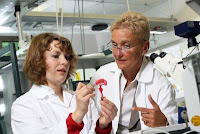
Artificial Liver for Drug Tests Thursday, 25 June 2009 If you have hay fever, headaches or a cold, it's only a short way to the nearest chemist. The drugs, on the other hand, can take eight to ten years to develop. Until now animal experiments have been an essential step, yet they continue to raise ethical issues. Dr. Johanna Schanz and Prof. Heike Mertsching (f.l.t.r.) work to develop an artificial liver. Credit: Fraunhofer/Dirk Mahler."Our artificial organ systems are aimed at offering an alternative to animal experiments," says Professor Heike Mertsching of the Fraunhofer Institute for Interfacial Engineering and Biotechnology IGB in Stuttgart. "Particularly as humans and animals have different metabolisms. 30 per cent of all side effects come to light in clinical trials." The test system, which Professor Mertsching has developed jointly with Dr. Johanna Schanz, should in future give pharmaceutical companies greater security and shorten the path to new drugs. Both researchers received the "Human-centered Technology" prize for their work. "The special feature, in our liver model for example, is a functioning system of blood vessels," says Dr. Schanz. "This creates a natural environment for cells." Traditional models do not have this, and the cells become inactive. "We don't build artificial blood vessels for this, but use existing ones – from a piece of pig's intestine." All of the pig cells are removed, but the blood vessels are preserved. Human cells are then seeded onto this structure – hepatocytes, which, as in the body, are responsible for transforming and breaking down drugs, and endothelial cells, which act as a barrier between blood and tissue cells. In order to simulate blood and circulation, the researchers put the model into a computer-controlled bioreactor with flexible tube pump, developed by the IGB. This enables the nutrient solution to be fed in and carried away in the same way as in veins and arteries in humans. "The cells were active for up to three weeks," says Dr. Schanz. "This time was sufficient to analyze and evaluate the functions. A longer period of activity is possible, however." The researchers established that the cells work in a similar way to those in the body. They detoxify, break down drugs and build up proteins. These are important pre-conditions for drug tests or transplants, as the effect of a substance can change when transformed or broken down – many drugs are only metabolized into their therapeutic active form in the liver, while others can develop poisonous substances. The researchers have demonstrated the basic possibilities for use of the tissue models – liver, skin, intestine and windpipe. At the moment, the test system is being examined. Within two years it could provide a safer alternative to animal experiments. ......... ZenMaster


For more on stem cells and cloning, go to CellNEWS at http://cellnews-blog.blogspot.com/ and http://www.geocities.com/giantfideli/index.html

Age Affects Function of Embryonic Muscle Stem Cell Genes

Adult satellite cells have distinct genetic requirements Thursday, 25 June 2009 Scientists working at the Carnegie Institution's Department of Embryology, with colleagues, have overturned previous research that identified critical genes for making muscle stem cells. It turns out that the genes that make muscle stem cells in the embryo are surprisingly not needed in adult muscle stem cells to regenerate muscles after injury. The finding challenges the current course of research into muscular dystrophy, muscle injury, and regenerative medicine, which uses stem cells for healing tissues, and it favours using age-matched stem cells for therapy. The study is published in the June 25 advance on-line edition of Nature. Previous studies have shown that two genes Pax3 and Pax7, are essential for making the embryonic and neonatal muscle stem cells in the mouse. Lead researcher Christoph Lepper, a predoctoral fellow in Carnegie's Chen-Ming Fan's lab and a Johns Hopkins student, for the first time looked at these two genes in promoting stem cells at varying stages of muscle growth in live mice after birth. As Christoph explained: "The paired-box genes, Pax3 and Pax7 are involved in the development of the skeletal muscles. It is well established that both genes are needed to produce muscle stem cells in the embryo. A previous student, Alice Chen, studied how these genes are turned on in embryonic muscle stem cells (also published in Nature). I thought that if they are so important in the embryo, they must be important for adult muscle stem cells. Using genetic tricks, I was able to suppress both genes in the adult muscle stem cells. I was totally surprised to find that the muscle stem cells are normal without them."


Mice muscle repair. This cross section of hind limb muscle tissue is from a mouse five days after injury. The uninjured cells are at top and stained red. The blue cells below are regenerating muscles cells. They were labelled with a blue stain and formed from muscle stem cells. Credit: Christoph Lepper.
The researchers then looked at whether the same was true upon injury, after which the repair process requires muscle stem cells to make new muscles. For this, they injured the leg muscles between the knee and ankle. They were again surprised that these muscle stem cells, without the two key embryonic muscle stem cell genes, could generate muscles as well as normal muscle stem cells. They even performed a second round of injury and found that the stem cells were still active. The scientists then wondered when these genes become unnecessary for muscle stem cells to regenerate muscles. It turned out that these embryonic genes are important to muscle stem cell creation up to the first three weeks after birth. What makes the muscle stem cells different after three weeks? The scientist believe that these two embryonic muscle stem cell genes also tell the stem cells to become quiet as the organism matures. After that time is reached, they "hand over" their jobs to a different set of genes. The researchers suggest that since the adult muscle stem cells are only activated when injury occurs (by trauma or exercise), they use a new set of genes from those used during embryonic development, which proceeds without injury. The scientists are eager to find these adult muscle stem cell genes. "We are just beginning to learn the basics of stem cell biology, and there are many surprises," remarked Allan Spradling, director of Carnegie's Department of Embryology. "This work illustrates the importance of carrying out basic research using animal models before rushing into the clinic with half-baked therapies." Reference: Adult satellite cells and embryonic muscle progenitors have distinct genetic requirements Christoph Lepper, Simon J. Conway & Chen-Ming Fan Nature advance online publication 25 June 2009, doi:10.1038/nature08209 ......... ZenMaster
For more on stem cells and cloning, go to
CellNEWS at http://cellnews-blog.blogspot.com/ and http://www.geocities.com/giantfideli/index.html

Tuesday 23 June 2009

Placenta: New Source for Harvesting Stem Cells

Children's Hospital Oakland scientists first to discover new source for harvesting stem cells Tuesday, 23 June 2009 A groundbreaking study conducted by Children's Hospital & Research Center at Oakland, California, is the first to reveal a new avenue for harvesting stem cells from a woman's placenta, or more specifically the discarded placentas of healthy newborns. The study also finds there are far more stem cells in placentas than in umbilical cord blood, and they can be safely extracted for transplantation. Furthermore, it is highly likely that placental stem cells, like umbilical cord blood and bone marrow stem cells, can be used to cure chronic blood-related disorders such as sickle cell disease, thalassaemia, and leukaemia. The study, led by Children's Hospital & Research Center Oakland scientists Frans Kuypers, PhD, and Vladimir Serikov, PhD, will be the feature story in the July 2009 issue of Experimental Biology and Medicine. The doctors and their team made the discoveries by harvesting term placentas from healthy women undergoing elective Caesarean sections. "Yes, the stem cells are there; yes, they are viable; and yes, we can get them out," declared Dr. Kuypers. Stem cells are essentially blank cells that can be transformed into any type of cell such as a muscle cell, a brain cell, or a red blood cell. Using stem cells from umbilical cord blood, Children's Hospital Oakland physicians have cured more than 100 kids with chronic blood-related diseases through their sibling donor cord blood transplantation program, which began in 1997. However, according to the American Cancer Society, each year at least 16,000 people with serious blood- related disorders are not able to receive the bone marrow or cord blood transplant they need because they cannot find a match. Dr. Kuypers explained that even when a patient receives a cord blood transplant, there may not be enough stem cells in the umbilical cord to successfully treat their disorder. Placentas, however, contain several times more stem cells than umbilical cord blood. "The greater supply of stem cells in placentas will likely increase the chance that an HLA (human leukocyte antigen) matched unit of stem cells engrafts, making stem cell transplants available to more people. The more stem cells, the bigger the chance of success," said Dr. Kuypers.


Chorionic villus of human term placenta. This is a microphotograph of chorionic villus of human term placenta immunostained for CD34 (Marker of endothelial and haematopoietic stem cells, red), CD31 (marker of endothelial cell, green) and nuclei (DAPI, blue). Non-endothelial CD34-positive cell is clearly observed in tissue of placenta. Credit: Society for Experimental Biology and Medicine.
In this report, said Dr. Serikov, we demonstrate for the first time that human placentas could provide abundant amounts of CD34+ CD133+ colony-forming cells, as well as other primitive hematopoietic progenitors, suitable for transplantation in humans. The total amount of live haematopoietic stem cells, or colony-forming units in culture that could be obtained from placentas was an order of magnitude larger than the number of hematopoietic stem cells obtained from cord blood from the same source. Haematopoietic stem cells which maintain their differentiation capacity, as well as stromal stem cells that support long-term culture of haematopoietic cells, can be harvested from perfusate of placenta following CXCR4 receptor blockade, said Dr. F. Kuypers. Importantly, live HPCs can similarly be obtained from whole cryopreserved placentas. Cells derived from placental tissue differentiated into all blood lineages in vitro. Animal experiments further demonstrated successful engraftment of placenta-derived HSC, which reconstituted haematopoiesis in immunodeficient mice. In summary, said Dr. F. Kuypers, our results indicate for the first time that human term placenta is a high capacity source of live and functional hematopoietic stem cells. By using placental circulation and stem cell receptor blockade an abundant amounts of hematopoietic stem cell could be easily obtained in sterile conditions by non-destructive methods. Dr. Steven R. Goodman, Editor-in-Chief of Experimental Biology and Medicine said "the outstanding importance of these results for practical haematology is determined by the fact that total number of stem cells that can be harvested from cord blood limits the efficacy of this stem cell source for transplants only to small children. These novel findings demonstrate that placenta may provide a source of autologous stem cells sufficient for reconstitution of haematopoiesis in adult patients. Use of methods to obtain haematopoietic cells from placenta, developed by Dr. Serikov and Dr. Kuypers as augmentation of cord blood-based therapy or replacement of bone marrow for transplantation will dramatically change whole field of transplantology." Drs. Kuypers and Serikov have also developed a patent-pending method that will allow placental stem cells to be safely harvested and made accessible for transplantation. The process involves freezing placentas in a way that allows them to later be defrosted and suffused with a compound that enables the extraction of viable stem cells. The method will make it possible for companies to gather, ship and store placentas in a central location. "We're looking for a partnership with industry to get placenta-derived stem cells in large quantities to the clinic," said Dr. Kuypers. He adds that much more research and grant funding are needed to explore the maximum potential of this latest discovery. He remains encouraged. "Someday, we will be able to save a lot more kids and adults from these horrific blood disorders." ......... ZenMaster
For more on stem cells and cloning, go to CellNEWS at http://cellnews-blog.blogspot.com/ and http://www.geocities.com/giantfideli/index.html

Thursday 18 June 2009

Researchers Edit Genes in Human Stem Cells

Researchers Edit Genes in Human Stem Cells Thursday, 18 June 2009 Researchers at the Johns Hopkins School of Medicine have successfully edited the genome of human- induced pluripotent stem cells, making possible the future development of patient-specific stem cell therapies. Reporting this week in Cell Stem Cell, the team altered a gene responsible for causing the rare blood disease paroxysmal nocturnal haemoglobinuria, or PNH, establishing for the first time a useful system to learn more about the disease. "To date, only about six genes have been successfully targeted or edited in human stem cells out of countless people and attempts — that's just not efficient enough if we want to move disease research and therapy forward," says Linzhao Cheng, Ph.D., an associate professor of gynaecology and obstetrics and member of the Johns Hopkins Institute of Cell Engineering. "We've been able to improve gene targeting and editing in human embryonic stem cells more than 200 fold." Cheng's lab and collaborators at Johns Hopkins study PNH, a condition where "friendly fire" kills patients' own blood cells and the body can't replenish the lost blood cells due to loss of normal blood stem cells. PNH is an acquired disease that occurs only in adults, according to Cheng. "It's a tough condition to study because we need to study it in blood stem cells and they're difficult to grow in the lab. So for years we've been trying to develop another cell system to better understand and perhaps fix what's going on in PNH." To establish a system for research, they used human embryonic stem cells which can be expanded unlimitedly in the laboratory, but they also had to create a mutation as found in a PNH patient. To target and remove the function of the one specific gene known to cause PNH, the research team improved on the standard approach of gene targeting, which can remove a functional gene or replace a dysfunctional gene. The gene targeting technology, first used successfully for mouse embryonic stem cells, won a Nobel Prize in Physiology or Medicine in 2007. Gene targeting exploits a cell's own ability to repair broken DNA. When DNA breaks from exposure to mutagens or other agents like DNA-cutting enzymes, DNA-repairing enzymes in the cell find and re-join the two exposed DNA ends. However, if another piece of DNA with exposed ends is floating around, it effectively can be spliced into the broken DNA during repair, and replace the defective copy. The team's technological improvement includes the use of custom-designed molecular scissors that are made by collaborators at Harvard University and University of Texas Southwestern Medical Center. These engineered DNA cutting enzymes make a precise break at specific locations in a cell's DNA — in this case in the gene that causes PNH. They added the molecular scissors and a fragment of DNA containing a gene that confers selection of rare targeted clones in both human embryonic stem cells and induced pluripotent stem cells. The latter, also known as iPS cells, are very similar to embryonic stem cells in biological properties, but generated by using adult tissues such as skin. Of all the cells surviving selection, they picked and grew eight iPS cell lines to study further, and five of those contained a targeted insertion at the gene site. Further examination showed that the cells contained the correct number of chromosomes, no longer contained any trace of the molecular scissors and had characteristics as cells from PNH patients that lack a group of cell surface molecules. "I commend my team, especially Dr. Jizhong Zou who spent three years with the help of many collaborators on this challenging project," says Cheng. "We're very excited about this accomplishment; it will enable better studies for other blood diseases. But there's still much to do before we can really use human iPS cells in clinical therapies." Cheng's team will continue to improve on techniques and begin applying these techniques to iPS cells from patients. Reference: Gene Targeting of a Disease-Related Gene in Human Induced Pluripotent Stem and Embryonic Stem Cells Jizhong Zou, Morgan L. Maeder, Prashant Mali, Shondra M. Pruett-Miller, Stacey Thibodeau-Beganny, Bin-Kuan Chou, Guibin Chen, Zhaohui Ye, In-Hyun Park, George Q. Daley, Matthew H. Porteus, J. Keith Joung, and Linzhao Cheng Cell Stem Cell, 18 June 2009, doi:10.1016/j.stem.2009.05.023 ......... ZenMaster


For more on stem cells and cloning, go to CellNEWS at http://cellnews-blog.blogspot.com/ and http://www.geocities.com/giantfideli/index.html

Fallopian Tubes Offer New Stem Cell Source

Fallopian Tubes Offer New Stem Cell Source Thursday, 18 June 2009 Human tissues normally discarded after surgical procedures could be a rich additional source of stem cells for regenerative medicine. New research from BioMed Central's open access Journal of Translational Medicine shows for the first time that human fallopian tubes are abundant in mesenchymal stem cells which have the potential of becoming a variety of cell types. It has previously been shown that mesenchymal stem cells obtained from umbilical cords, dental pulp and adipose tissue, which are all biological discards, are able to differentiate into muscle, fat, bone and cartilage cell lineages; therefore, the search for sources to obtain multipotent stem cells from discarded tissues and without ethical problems is of great interest. Tatiana Jazedje, and the research team from Human Genome Research Centre at the University of São Paulo, directed by Mayana Zatz, with the collaboration of medical doctors from the reproductive area, set out to isolate and assess the differentiation potential of mesenchymal stem cells from discarded human fallopian tubes. In the study, human fallopian tubes were obtained from hysterectomy and other gynaecological procedures from fertile women in their reproductive years (range 35-53 years) who had not undergone hormonal treatment for at least three months prior to surgery. The Brazilian team found that human fallopian tube mesenchymal stem cells could be easily isolated and expanded in vitro, and are able to differentiate into muscle, fat, cartilage and bone cell lines. The cells' chromosome complement showed no abnormalities, suggesting chromosomal stability. "In addition to providing an additional potential source for regenerative medicine, these findings might contribute to reproductive science as a whole," Jazedje commented. "Moreover, the use of human tissue fragments that are usually discarded in surgical procedures does not pose ethical problems," Jazedje concluded. Reference: Human fallopian tube: a new source of multipotent adult mesenchymal stem cells discarded in surgical procedures Tatiana Jazedje, Paulo M Perin, Carlos E Czeresnia, Mariangela Maluf, Silvio Halpern, Mariane Secco, Daniela F Bueno, Natassia M Vieira, Eder Zucconi and Mayana Zatz Journal of Translational Medicine (in press) ......... ZenMaster


For more on stem cells and cloning, go to CellNEWS at http://cellnews-blog.blogspot.com/ and http://www.geocities.com/giantfideli/index.html

Tuesday 16 June 2009

Gene Vital to Early Embryonic Cells Forming a Normal Heart and Skull

Gene Vital to Early Embryonic Cells Forming a Normal Heart and Skull Tuesday, 16 June 2009 New research from Cincinnati Children's Hospital Medical Center highlights the critical role a certain gene and its protein play during early embryonic development on formation of a normal heart and skull. In a study posted online June 15 by the Proceedings of the National Academy of Sciences, a research team at Cincinnati Children's reports that too little of the gene/protein SHP2 interferes with the normal developmental activity of what are called neural crest cells. These cells, which occur very early in embryonic development, migrate to specific regions of the embryo. While doing so, the cells are supposed to differentiate and give rise to certain nerve tissues, craniofacial bones or smooth muscle tissue of the heart. "Our findings show that a deficiency of SHP2 in neural crest cells results in a failure of cell differentiation at diverse sites in the developing embryo," said Jeffrey Robbins, Ph.D., co-director of the Heart Institute at Cincinnati Children's and senior investigator of the study. "This leads to anatomical and functional deficits so severe that it precludes viability of the developing foetus." SHP2 is a tyrosine phosphatase – an enzyme that helps trigger a cascade of biochemical reactions in cells as they specify to form certain tissues. Although the study was conducted using mouse embryos, the findings are significant in efforts to understand congenital malformations of the heart and craniofacial region in people. Especially relevant, the researchers said, is the insight gained into early molecular events during embryonic development that might help explain such birth defects. Dr. Robbins said the findings from this study could be used to develop specific drugs that could target the affected pathway, leading to treatment of heart and craniofacial malformations. About 4 percent of human infants are born with congenital malformations. Abnormal heart development is the most common human birth defect, affecting about 1 percent of newborns. The researcher team also wants to explore the exact alterations in neural crest cell migration, expansion and differentiation that contribute to birth defects of other organ systems. ......... ZenMaster


For more on stem cells and cloning, go to CellNEWS at http://cellnews-blog.blogspot.com/ and http://www.geocities.com/giantfideli/index.html

Monday 15 June 2009

Huntington's disease Deciphered

Mutated huntingtin activates a neuron specific kinase called JNK3 Monday, 15 June 2009 Researchers at the University of Illinois at Chicago College of Medicine have discovered how the mutated huntingtin gene acts on the nervous system to create the devastation of Huntington's disease. The report of their findings is available in Nature Neuroscience online. The researchers were able to show that the mutated huntingtin gene activates a particular enzyme, called JNK3, which is expressed only in neurons and, further, to show what effect activation of that enzyme has on neuron function. Huntington's disease is an adult onset neurodegenerative disease marked by progressive mental and physical deterioration. It has been known for more than a decade that everyone who develops the disease has mutations in a particular gene, called huntingtin, according to Scott Brady, professor and head of anatomy and cell biology at the UIC College of Medicine. "There are several puzzling aspects of this disease," said Brady, who is co-principal investigator on the study. "First, the mutation is there from day one. How is it that people are born with a perfectly functioning nervous system, despite the mutation, but as they grow up into their 30s and 40s they start to develop these debilitating symptoms? We need to understand why the protein is bad at 40 but it wasn't bad at 4." The second problem, according to Brady, is that the gene is expressed not just in the nervous system but in other parts of the body. However, the only part of the body that is affected is the nervous system. Why are neurons being affected? Brady, Gerardo Morfini, assistant professor of anatomy and cell biology at UIC and co-principal investigator of the study, and their colleagues began looking for a mechanism that could explain all the pieces of the puzzle. They found that at extremely low concentrations, huntingtin was a potent inhibitor of axonal transport, the system within the neuron that shuttles proteins from the cell body where they are synthesized to the synaptic terminals where they are needed. A neuron's critical role in making connections may require it to make the cellular trunk, called an axon, between the cell body and the synaptic terminal to be very long. Some cells have axons that reach half the body's length – for a tall person, a meter or more. But even in the brain, axonal projections are very long compared to other cells. In addition to the challenge of distance, neurons are very complex cells with many specialized areas necessary to carry out synaptic connections, requiring a robust transport system. "Inhibition of neuronal transport is enough to explain what is happening in Huntington's," said Brady. Loss of delivery of materials to the terminals results in loss of transmission of signals from the neuron. Loss of signal transmission causes the neurons to begin to die back, leading to reduced transmissions, more dying back and eventual neuronal cell death. This mechanism also explains the late onset of the disease, Brady said. Activation of JNK3 reduces transport but does not eliminate it. Young neurons have a robust transport system, but transport gradually declines with age. "If you take a hit when you're very young, you still are making more and transporting more proteins in each neuron than you need," Brady said. "But as you get older and older, the neuron produces and transports less. Each hit diminishes the system further. Eventually, the neuron falls below the threshold needed to maintain cell health." Brady's group has also linked this pattern of progressive neurodegeneration – marked by a loss of signalling between neurons, a slow dying back of neurons, and eventual neuron death – to damage to the transport system in several other hereditary adult-onset neurodegenerative diseases and to Alzheimer's disease. "There is a common theme and a common Achilles heel of the neuron that underlies all these diseases," Brady said. "We've invented a word, dysferopathy, (from the Greek 'fero', to carry or transport) for these adult-onset neurodegenerative diseases. All have disruption of the axonal transport system in common." Reference: Pathogenic huntingtin inhibits fast axonal transport by activating JNK3 and phosphorylating kinesin Gerardo A Morfini, Yi-Mei You, Sarah L Pollema, Agnieszka Kaminska, Katherine Liu, Katsuji Yoshioka, Benny Björkblom, Eleanor T Coffey, Carolina Bagnato, David Han, Chun-Fang Huang, Gary Banker, Gustavo Pigino & Scott T Brady Nature Neuroscience Published online: 14 June 2009, doi:10.1038/nn.2346 ......... ZenMaster


For more on stem cells and cloning, go to CellNEWS at http://cellnews-blog.blogspot.com/ and http://www.geocities.com/giantfideli/index.html

Sperm Delivers More Complex Material than Thought

New study explores dad's role in shaping a healthy baby Monday, 15 June 2009 It was long believed that conception does not involve a meeting of equals. The egg is a relatively large, impressive biological factory compared with the tiny sperm, which delivers to the egg one copy of the father’s genes. However, a new study from Huntsman Cancer Institute (HCI) at the University of Utah reveals that the father’s sperm delivers much more complex genetic material than previously thought. The findings could lead to a diagnostic test to help couples deal with infertility. Researchers discovered particular genes packaged in a special way within the sperm, and that may promote the development of the foetus. “Our findings show that the father plays an active role in packaging his genome to help ensure a healthy baby,” says study co-leader Brad Cairns, Ph.D., investigator with HCI and the Howard Hughes Medical Institute, and professor of oncological sciences at the University of Utah. “However, they also raise the possibility that a man’s aging, health and lifestyle may alter this packaging and negatively affect fertility and embryo development.” During foetal development, certain genes make decisions about organ and tissue development. The new research shows that in sperm, these genes are wrapped in special packaging materials called ‘modified histones.’ These modified histones appear to be key factors in ensuring genes are activated or repressed at the right level, place and time, which helps the fertilized egg develop properly, known as epigenetic inheritance. Chromosomes are long strands of DNA containing thousands of genes, and their packaging helps determine which genes turn on and off. Understanding how these genes are activated or repressed leads to a better understanding of how disorders like birth defects and cancer develop. “Genes have on-and-off switches, and understanding them allows us to target them, leading to possible treatments, cures or prevention strategies,” says Cairns. “That’s the good news.” An implication of this study is that factors such as genetic mutations, age or lifestyle may affect sperm chromosome packaging, leading to infertility. “We are hopeful that this work will soon lead to a clinical diagnostic test that will help couples with infertility problems make better informed decisions regarding their prospects for a healthy child. We will also be testing if aspects of a man’s lifestyle – such as age, diet or health – affect proper packaging and fertility,” says Cairns. Other future work includes how decision-making genes are packaged in eggs, which remains a major mystery. The study is set for publication June 14 in the online edition of the journal Nature. The research involved collaboration between Cairns’ lab at HCI and the University of Utah’s in vitro fertilization (IVF) and andrology lab led by Doug Carrell, along with their joint graduate student, Sue Hammoud. About Huntsman Cancer Institute: Huntsman Cancer Institute (HCI) at the University of Utah marks its 10th anniversary in 2009. HCI was founded by Jon M. Huntsman to fulfil his dream of finding a cure for cancer through genetic research. In the last 10 years, HCI has grown to become one of America’s major cancer research centres. HCI is part of the University of Utah Health Care system and is ranked consistently by U.S. News & World Report as one of the top cancer hospitals in the country. Reference: Distinctive chromatin in human sperm packages genes for embryo development Saher Sue Hammoud, David A. Nix, Haiying Zhang, Jahnvi Purwar, Douglas T. Carrell & Bradley R. Cairns Nature advance online publication 14 June 2009, doi:10.1038/nature08162 ......... ZenMaster


For more on stem cells and cloning, go to CellNEWS at http://cellnews-blog.blogspot.com/ and http://www.geocities.com/giantfideli/index.html

Tuesday 9 June 2009

Batten Disease and Stem Cells Treatment Clinical Trial Update

StemCells Inc. Announces Positive Phase I Batten Trial Results Monday, 08 June 2009 StemCells Inc. announced today positive results from the first Phase I clinical trial of its proprietary HuCNS-SC® product candidate (purified human neural stem cells), including demonstration of a favourable safety profile along with evidence of engraftment and long-term survival of the HuCNS-SC cells. The Phase I trial was designed primarily to assess the safety of HuCNS-SC cells as a potential cell-based therapeutic. Six patients with advanced stages of infantile and late infantile neuronal ceroid lipofuscinosis (NCL), often referred to as Batten disease, were transplanted with HuCNS-SC cells and followed for 12 months. Overall, the Phase I data demonstrated that high doses of HuCNS-SC cells, delivered by a direct transplantation procedure into multiple sites within the brain, followed by twelve months of immunosuppression, were well tolerated by all six patients enrolled in the trial. The patients’ medical, neurological and neuropsychological conditions, following transplantation, appeared consistent with the normal course of the disease. The independent Data Monitoring Committee (DMC), a multi-disciplinary group of experts in neurosurgery, transplant medicine, genetics, and neurology responsible for overseeing the safety of the trial, has also concurred with the Company’s assessment of the safety profile of the test product and procedure. The trial was conducted at Oregon Health & Science University (OHSU) Doernbecher Children's Hospital and was completed in January 2009. StemCells will present the final study report to the FDA and plans to pursue future clinical development of HuCNS-SC as a potential treatment for infantile and late infantile NCL. "We are very pleased and encouraged by the results of this landmark trial,” said Martin McGlynn, president and chief executive officer of StemCells. "As this was the first-ever FDA-authorized study of human neural stem cells as a potential therapeutic agent in humans, the favourable data we obtained is especially meaningful. Completing this first trial also marked an important milestone in the evolution of our cell-based product candidates from research and development to human clinical studies. We are deeply grateful for the support of the patients’ families who enabled us to make an important advance in our search for a therapy that might one day benefit not only children with Batten disease, but also those suffering from other serious neurodegenerative diseases.” Commenting on the trial data, Stephen Huhn, MD, FACS, FAAP, vice president and head of the Company’s CNS Program, stated: "The HuCNS-SC cells were well tolerated even at very high dose levels – as many as one billion cells were transplanted into certain patients. Given the considerable number of cells transplanted, together with the very fragile nature of the patients involved, the positive safety data we observed is particularly noteworthy.” StemCells previously reported the loss of the second patient enrolled in the trial, who died from the natural progression of the disease approximately one year post-transplant. Because the family consented to an autopsy examination of the brain, the Company was able to establish that the donor cells had engrafted and survived, despite severe brain atrophy related to the NCL. By permitting the autopsy, the family allowed the researchers to learn very important details that will potentially benefit future patients. "Our strategy for these lysosomal storage diseases is to protect the patient’s remaining neurons by transplanting donor cells without the genetic defect that causes NCL into the brain,” continued Dr. Huhn. "These healthy neural stem cells have the potential to produce the enzyme currently lacking for proper function and survival of the patient’s brain cells. In this first trial, however, the patients already had a severe amount of neuronal degeneration and brain atrophy due to the advanced stage of their disease and only a limited number of brain cells remaining to protect, making it difficult to measure any degree of efficacy. Our interpretation of potential efficacy measurements was also limited by the number of subjects enrolled in the trial and the absence of a control group. Consequently, now that we have demonstrated a favourable safety profile and evidence of long term donor cell survival, our objective is to initiate a second trial designed to test the potential for efficacy in patients in a much earlier stage of the disease.” Robert Steiner, MD, FAAP, FACMG, co-principal investigator, professor of paediatrics and molecular and medical genetics, and vice chairman for paediatric research at OHSU Doernbecher Children's Hospital, stated: "The OHSU research team worked very hard to carry out this highly complex research and is heartened to see that this approach appears to be safe. We are delighted that this first trial of human neural stem cells was successful and offers some hope for effective treatment of NCL and other neurodegenerative disorders.” "It was a privilege for our team to care for these precious children,” added Nathan Selden, MD, Ph.D., FACS, FAAP, co-principal investigator, Campagna Associate Professor and head, division of paediatric neurological surgery at OHSU Doernbecher Children’s Hospital. "We are indebted to our patients and their families for taking us into this new era of therapy for the central nervous system. We hold out great hope in the future for them and for others around the world with similar diseases that today have no cure.” Trial Design The Phase I trial was designed primarily to assess the safety of HuCNS-SC cells as a potential treatment for infantile and late infantile NCL, including the tolerability of multiple interventions (surgery, immunosuppression and the HuCNS-SC cells). Six patients with either infantile or late infantile NCL were enrolled in the open-label, dose-escalating Phase I study and transplanted with HuCNS-SC cells. Enrolment in the trial was limited to those patients in advanced stages of the disease with significant neurological and cognitive impairment (patients whose developmental age was demonstrated to be less than two-thirds of their chronological age). Two dose levels were administered, with the first three patients receiving a target dose of approximately 500 million cells, and the other three patients receiving a target dose of approximately one billion cells. The HuCNS-SC cells were directly transplanted into each patient’s brain via a neurosurgical procedure, and patients were immuno-suppressed for 12 months following transplantation. The patients were evaluated and assessed at regular intervals using a comprehensive range of medical, neurological and neuropsychological tests, both before transplantation to establish a baseline, and over the course of 12 months following transplantation. Following completion of the Phase I trial, the patients were automatically enrolled in a separate four-year follow-up study. Summary of Data The most common non-serious adverse events observed during the trial were related to immunosuppression. A total of 13 serious adverse events were noted, of which 54% were reported for one patient, and none of which were considered related to the HuCNS-SC cells. Magnetic resonance images (MRIs) of each patient’s brain were taken at baseline, immediately following surgery, and at six months and 12 months following transplantation to evaluate the injection sites. Of the 48 total injection sites (eight per patient), no MRI abnormalities related to the cells were detected. A single artefact at one transplant site in one patient was evident by MRI, and was considered a minor, harmless change related to the surgery. The previously reported death of one patient approximately one year following transplantation was determined, after an autopsy and a review of medical records in consultation with the DMC, to be the result of the natural progression of the disease. The evidence of regional engraftment and survival of the HuCNS-SC cells from this autopsy supports continued effort toward the goal of demonstrating efficacy. About Neuronal Ceroid Lipofuscinosis (Batten Disease) Neuronal ceroid lipofuscinosis (NCL) is a fatal neurodegenerative disorder that afflicts infants and young children. The disorder, often referred to as Batten disease, is caused by genetic mutations, and children who inherit the defective gene are unable to produce enough of an enzyme that processes cellular waste substances that accumulate in a part of cells known as the lysosome. Without the enzyme, the cellular waste builds up, and eventually the cells cannot function and die. Children with NCL appear healthy when born, but as their brain cells die, they begin to suffer seizures and progressively lose motor skills, sight and mental capacity. Eventually, they become blind, bedridden and unable to communicate or function independently. There currently is no cure for the disease. The infantile and late infantile forms of NCL are caused by different genetic mutations. As the names imply, the two forms begin to afflict patients at different stages of infancy, but both have similar disease progression and outcomes. About HuCNS-SC® Cells StemCells’ lead product candidate, HuCNS-SC cells, is a purified composition of normal human neural stem cells that are expanded and stored as banks of cells. The Company’s preclinical research has shown that HuCNS-SC cells can be directly transplanted; they engraft, migrate, differentiate into neurons and glial cells; and they survive for as long as one year with no sign of tumour formation or adverse effects. These findings show that HuCNS-SC cells, when transplanted, act like normal stem cells, suggesting the possibility of a continual replenishment of normal human neural cells. About StemCells Inc. StemCells, Inc. is a clinical-stage biotechnology company focused on the research, development and commercialization of products derived from stem cell technologies. In its therapeutic product development programs, StemCells is focused on developing cell-based therapeutics to treat diseases of the central nervous system and liver. StemCells has pioneered the discovery and development of HuCNS-SC® cells, its highly purified, expandable population of human neural stem cells. StemCells has completed a six-patient Phase I clinical trial of its proprietary HuCNS-SC product candidate as a treatment for neuronal ceroid lipofuscinosis (NCL), a rare and fatal neurodegenerative disease that affects infants and young children. StemCells has also received approval from the Food and Drug Administration (FDA) to initiate a Phase I clinical trial of the HuCNS-SC cells to treat Pelizaeus-Merzbacher Disease (PMD), a rare and fatal brain disorder that mainly affects young children. StemCells, through its wholly owned subsidiaries Stem Cell Sciences UK Ltd and Stem Cell Sciences Australia Pty, is also pursuing applications of its cell-based technologies to develop research tools, such as cell-based assays, media and reagent tools, which the Company believes represent nearer-term commercial opportunities. StemCells has exclusive rights to approximately 55 issued or allowed U.S. patents and approximately 200 granted or allowed non-US patents. Further information about StemCells is available on its web site at: www.stemcellsinc.com. About Oregon Health & Science University Doernbecher Children’s Hospital OHSU is the state's only health and research university and Oregon's only academic health centre. OHSU is Portland's largest employer and the fourth largest in Oregon (excluding government). OHSU's size contributes to its ability to provide many services and community support activities not found anywhere else in the state. It serves patients from every corner of the state, and is a conduit for learning for more than 3,400 students and trainees. OHSU is the source of more than 200 community outreach programs that bring health and education services to every county in the state. OHSU Doernbecher Children's Hospital is a world-class facility that each year cares for tens of thousands of children from Oregon, southwest Washington and around the nation, including national and international referrals for specialty care. Children have access to a full range of paediatric care, not just treatments for serious illness or injury, resulting in more than 120,000 outpatient visits, discharges, surgeries and paediatric transports annually. In addition, nationally recognized physicians ensure that children receive exceptional care at OHSU Doernbecher, including outstanding cancer treatment, specialized neurology care and highly sophisticated heart surgery in the most patient- and family-centred environment. Paediatric experts from OHSU Doernbecher travel throughout Oregon and southwest Washington to provide specialty care to some 2,800 children at more than 154 outreach clinics in 13 locations. ......... ZenMaster


For more on stem cells and cloning, go to CellNEWS at http://cellnews-blog.blogspot.com/ and http://www.geocities.com/giantfideli/index.html

Thursday 4 June 2009

Another Chinese Group Create Pig Embryonic-like Stem Cells

Engineered pig stem cells bridge the mouse-human gap Thursday, 04 June 2009 The discovery that adult skin cells can be 'reprogrammed' to behave like stem cells has been a major scientific boon, providing a way to tap the potential of embryonic stem cells without the associated ethical quandaries. Now, in a study appearing online in JBC, researchers have created a line of such reprogrammed stem cells from adult pigs. As pigs are large animals with a physiology very similar to humans, this work provides a valuable model to study the therapeutic potential of this new "induced pluripotent stem cell" (iPS) technology. iPS cells have already been developed from both mice and humans. Both systems will help researchers answer many biological and genetic questions about these cells, but still leave a gap before clinical applications can begin. These iPS cells cannot be tested on humans before thorough safety and efficacy trials in animal models, but the size, physiology and short lifespan of mice makes them less than ideal for these trials. Duanqing Pei and colleagues, from South China Institute for Stem Cell Biology and Regenerative Medicine, Guangzhou Institutes of Biomedicine and Health, China, turned to a better pre-clinical model: pigs. These large animals share a remarkably similar biology to humans, as evidenced by their already extensive contributions to medicine, such as using pig insulin to treat diabetes or pig heart valves in transplant surgery. The research group modified the current iPS protocols to successfully generate a line of stem cells from a miniature Tibetan pig (whose smaller size would make breeding and maintenance easier). A biochemical analysis revealed these cells expressed the key proteins that would classify them as 'stem cells' and had the ability to differentiate into many other types of cells. Importantly, these pig iPS cells more closely resembled human stem cells than other animals, confirming their value in pre-clinical studies. The researchers believe porcine iPS technology is an emerging and exciting field that should progress quickly and lead to many applications. Reference: Generation of induced pluripotent stem cell lines from Tibetan miniature pig Miguel Angel Esteban, Jianyong Xu, Jiayin Yang, Meixiu Peng, Dajiang Qin, Wen Li, Zhuoxin Jiang, Jiekai Chen, Kang Deng, Mei Zhong, Jinglei Cai, Liangxue Lai, and Duanqing Pei Journal of Biological Chemistry, April 17, 2009, doi:10.1074/jbc.M109.008938 ......... ZenMaster


For more on stem cells and cloning, go to CellNEWS at http://cellnews-blog.blogspot.com/ and http://www.geocities.com/giantfideli/index.html

Wednesday 3 June 2009

Small Molecules Mimic Natural Gene Regulators

Small Molecules Mimic Natural Gene Regulators Wednesday, 03 June 2009 In the quest for new approaches to treating and preventing disease, one appealing route involves turning genes on or off at will, directly intervening in ailments such as cancer and diabetes, which result when genes fail to turn on and off as they should. Scientists at the University of Michigan and the University of California at Berkeley have taken a step forward on that route by developing small molecules that mimic the behaviour and function of a much larger and more complicated natural regulator of gene expression. The research, by associate professor of chemistry Anna Mapp and co-workers, is described in the current issue of the journal ACS Chemical Biology. Molecules that can prompt genes to be active are called transcriptional activators because they influence transcription — the first step in the process through which instructions coded in genes are used to produce proteins. Transcriptional activators occur naturally in cells, but Mapp and other researchers have been working to develop artificial transcription factors (ATFs) — non-natural molecules programmed to perform the same function as their natural counterparts. These molecules can help scientists probe the transcription process and perhaps eventually be used to correct diseases that result from errors in gene regulation. In previous work, Mapp and co-workers showed that an ATF they developed was able to turn on genes in living cells, but they weren't sure it was using the same mechanism that natural activators use. Both natural transcriptional activators and their artificial counterparts typically have two essential parts: a DNA-binding domain that homes in on the specific gene to be regulated, and an activation domain that attaches itself to the cell's machinery through a key protein-to-protein interaction and spurs the gene into action. The researchers wanted to know whether their ATFs attached to the same sites in the transcriptional machinery that natural activators did. In the current work, the team showed that their ATFs bind to a protein called CBP, which interacts with many natural activators, and that the specific site where their ATFs bind is the same site utilized by the natural activators, even though the natural activators are much larger and more complex. Then the researchers altered their ATFs in various ways and looked to see how those changes affected both binding and ability to function as transcriptional activators. Any change that prevented an ATF from binding to CBP also prevented it from doing its job. This suggests that, for ATFs as for natural activators, interaction with CBP is key to transcriptional activity. "Taken together, the evidence suggests that the small molecules we have developed mimic both the function and the mechanism of their natural counterparts," said Mapp, who has a joint appointment in the College of Pharmacy's Department of Medicinal Chemistry. Next the researchers want to understand in more detail exactly how the small molecules bind to that site. "Then we'll use that information to design better molecules." Reference: Amphipathic Small Molecules Mimic the Binding Mode and Function of Endogenous Transcription Factors Sara J. Buhrlage, Caleb A. Bates, Steven P. Rowe, Aaron R. Minter, Brian B. Brennan, Chinmay Y. Majmudar, David E. Wemmer, Hashim Al-Hashimi and Anna K. Mapp ACS Chem. Biol., 2009, 4 (5), pp 335–344, DOI: 10.1021/cb900028j ......... ZenMaster


For more on stem cells and cloning, go to CellNEWS at http://cellnews-blog.blogspot.com/ and http://www.geocities.com/giantfideli/index.html

Chinese Scientists Create Pig Embryonic-like Stem Cells

Discovery has far-reaching implications for animal and human health Wednesday, 03 June 2009 Scientists have managed to induce cells from pigs to transform into pluripotent stem cells – cells that, like embryonic stem cells, are capable of developing into any type of cell in the body. It is the first time in the world that this has been achieved using somatic cells (cells that are not sperm or egg cells) from any animal with hooves (known as ungulates). The implications of this achievement are far-reaching; the research could open the way to creating models for human genetic diseases, genetically engineering animals for organ transplants for humans, and for developing pigs that are resistant to diseases such as swine flu. The work is the first research paper to be published online today (Wednesday 3 June) in the newly launched Journal of Molecular Cell Biology. Dr Lei Xiao, who led the research, said: "To date, many efforts have been made to establish ungulate pluripotent embryonic stem cells from early embryos without success. This is the first report in the world of the creation of domesticated ungulate pluripotent stem cells. Therefore, it is entirely new, very important and has a number of applications for both human and animal health." Dr Xiao, who heads the stem cell lab at the Shanghai Institute of Biochemistry and Cell Biology, Shanghai, China, and colleagues succeeded in generating induced pluripotent stem cells by using transcription factors to re-programme cells taken from a pig's ear and bone marrow. After the cocktail of reprogramming factors had been introduced into the cells via a virus, the cells changed and developed in the laboratory into colonies of embryonic-like stem cells. Further tests confirmed that they were, in fact, stem cells capable of differentiating into the cell types that make up the three layers in an embryo – endoderm, mesoderm and ectoderm – a quality that all embryonic stem cells have. The information gained from successfully inducing pluripotent stem cells (iPS cells) means that it will be much easier for researchers to go on to develop embryonic stem cells (ES cells) that originate from pig or other ungulate embryos. Dr Xiao said: "Pig pluripotent stem cells would be useful in a number of ways, such as precisely engineering transgenic animals for organ transplantation therapies. The pig species is significantly similar to humans in its form and function, and the organ dimensions are largely similar to human organs. We could use embryonic stem cells or induced stem cells to modify the immune-related genes in the pig to make the pig organ compatible to the human immune system. Then we could use these pigs as organ donors to provide organs for patients that won't trigger an adverse reaction from the patient's own immune system.” "Pig pluripotent stem cell lines could also be used to create models for human genetic diseases. Many human diseases, such as diabetes, are caused by a disorder of gene expression. We could modify the pig gene in the stem cells and generate pigs carrying the same gene disorder so that they would have a similar syndrome to that seen in human patients. Then it would be possible to use the pig model to develop therapies to treat the disease.” "To combat swine flu, for instance, we could make a precise, gene-modified pig to improve the animal's resistance to the disease. We would do this by first, finding a gene that has anti-swine flu activity, or inhibits the proliferation of the swine flu virus; second, we can introduce this gene to the pig via pluripotent stem cells – a process known as gene 'knock-in'. Alternatively, because the swine flu virus needs to bind with a receptor on the cell membrane of the pig to enter the cells and proliferate, we could knock out this receptor in the pig via gene targeting in the pig induced pluripotent stem cell. If the receptor is missing, the virus will not infect the pig." In addition to medical applications for pigs and humans, Dr Xiao said his discovery could be used to improve animal farming, not only by making the pigs healthier, but also by modifying the growth-related genes to change and improve the way the pigs grow. However, Dr Xiao warned that it could take several years before some of the potential medical applications of his research could be used in the clinic. The next stage of his research is to use the pig iPS cells to generate gene-modified pigs that could provide organs for patients, improve the pig species or be used for disease resistance. The modified animals would be either "knock in" pigs where the iPS or ES cells have been used to transfer an additional bit of genetic material (such as a piece of human DNA) into the pig's genome, or "knock out" pigs where the technology is used to prevent a particular gene functioning. Commenting on the study, the journal's editor-in-chief, Professor Dangsheng Li, said: "This research is very exciting because it represents the first rigorous demonstration of the establishment of pluripotent stem cell in ungulate species, which will open up interesting opportunities for creating precise, gene-modified animals for research, therapeutic and agricultural purposes." This release is available in Chinese: 世界首次:中国科学家培育出猪的干细胞 该发现对于动物和人类健康具有深远影响 Reference: Generation of pig induced pluripotent stem cells with a drug-inducible system Journal of Molecular Cell Biology. doi:10.1093/jmcb/jmp003 ......... ZenMaster


For more on stem cells and cloning, go to CellNEWS at http://cellnews-blog.blogspot.com/ and http://www.geocities.com/giantfideli/index.html

Monday 1 June 2009

Stem Cell Protein Offers a New Cancer Target

LIN28, which maintains cell 'stemness,' is abundant in advanced cancers and transforms cells to cancerous state Monday, 01 June 2009 A protein abundant in embryonic stem cells is now shown to be important in cancer, and offers a possible new target for drug development, report researchers from the Stem Cell Program at Children's Hospital Boston. Last year, George Daley, MD, PhD, and graduate student Srinivas Viswanathan, in collaboration with Richard Gregory, PhD, also of the Stem Cell Program at Children's, showed that the protein LIN28 regulates an important group of tumour-suppressing microRNAs known as let-7. Increasing LIN28 production in a cell prevented let-7 from maturing, making the cell more immature and stem-like. Since these qualities also make a cell more cancerous, and because low levels of mature let-7 have been associated with breast and lung cancer, the discovery suggested that LIN28 might be oncogenic. Now, publishing Advance Online in Nature Genetics on May 31, Daley, Viswanathan and colleagues show directly that LIN28 can transform cells to a cancerous state, and that it is abundant in a variety of advanced human cancers, particularly liver cancer, ovarian cancer, chronic myeloid leukaemia, germ cell tumours and Wilm's tumour (a childhood kidney cancer). They believe that overall, LIN28 and a related protein, LIN28B, may be involved in some 15 percent of human cancers. By blocking or suppressing LIN28, it might be possible to revive the let-7 family's natural tumour-suppressing action. "Linking this protein to advanced cancer is a very exciting new result," says Daley, Director of Stem Cell Transplantation at Children's, and also affiliated with Children's Division of Hematology/Oncology, the Dana-Farber Cancer Institute and the Harvard Stem Cell Institute. "It gives us a new target to attack, especially in the most resistant and hard-to-treat cases." LIN28, which is abundant in embryonic stem cells and prevents them from differentiating into specific cell types, was originally discovered to influence embryonic development in worms some 25 years ago. Development, stem cell generation and carcinogenesis are known to be closely related, but until last year's study connected LIN28 to let-7, it had not been clear how. "LIN28 is a fascinating protein that acts both in stem cells and cancers, and is teaching us that cancer is often a disease of stem cells," says Daley. Viswanathan, Daley and colleagues are busily searching for ways to inhibit LIN28, which could provide promising new drugs for advanced cancer. Reference: Lin28 promotes transformation and is associated with advanced human malignancies Srinivas R Viswanathan, John T Powers, William Einhorn, Yujin Hoshida, Tony L Ng, Sara Toffanin, Maureen O'Sullivan, Jun Lu, Letha A Phillips, Victoria L Lockhart, Samar P Shah, Pradeep S Tanwar, Craig H Mermel, Rameen Beroukhim, Mohammad Azam, Jose Teixeira, Matthew Meyerson, Timothy P Hughes, Josep M Llovet, Jerald Radich, Charles G Mullighan, Todd R Golub, Poul H Sorensen & George Q Daley Nature Genetics, Published online: 31 May 2009, doi:10.1038/ng.392 ......... ZenMaster


For more on stem cells and cloning, go to CellNEWS at http://cellnews-blog.blogspot.com/ and http://www.geocities.com/giantfideli/index.html