Monday 21 December 2009

Lack of Diversity in Embryonic Stem Cell Lines

Lack of Diversity in Embryonic Stem Cell Lines Monday, 21 December 2009 The most widely used human embryonic stem cell lines lack genetic diversity, a finding that raises social justice questions that must be addressed to ensure that all sectors of society benefit from stem cell advances, according to a University of Michigan research team. In the first published study of its kind, the U-M team analyzed 47 embryonic stem cell lines, including most of the lines commonly used by stem cell researchers. The scientists determined the genetic ancestry of each line and found that most were derived from donors of northern and western European ancestry. Several of the lines are of Middle Eastern or southern European ancestry. Two of the lines are of East Asian origin. None of the lines were derived from individuals of recent African ancestry, from Pacific Islanders, or from populations indigenous to the Americas. In addition, U-M researchers identified several instances in which more than one cell line came from the same embryo donors, further reducing the overall genetic diversity of the most widely available lines. "Embryonic stem cell research has the potential to change the future of medicine," said Sean Morrison, director of the U-M Center for Stem Cell Biology and one of the study leaders. "But there's a lack of diversity among today's most commonly used human embryonic stem cell lines, which highlights an important social justice issue." "We expected Europeans to be overrepresented, but we were surprised by how little diversity there is," he said. For the study, Morrison teamed up with two colleagues at the U-M Life Sciences Institute: stem cell scientist Jack Mosher and population geneticist Noah Rosenberg. Their findings are scheduled to be published online Wednesday in the New England Journal of Medicine. A fundamental principle of medical research is that new therapies are tested on patients that mirror the diversity in society, because certain groups may respond to medications and treatments differently. By evaluating new therapies in diverse patients, researchers are more likely to detect the different effects these therapies might have. Embryonic stem cell lines are being used to develop new cellular therapies for spinal cord injuries and various diseases, to screen for new drugs and to better understand inherited diseases. It is crucial that diverse lines are available for this research to ensure that all patients benefit from the results, Morrison said. "If that's not done, we run the risk of leaving certain groups in our society behind," said Morrison, who is a Howard Hughes Medical Institute investigator at U-M. The U-M report comes as Michigan researchers launch new projects made possible by a recent state constitutional amendment allowing researchers in the state to derive new human embryonic stem cell lines using approaches already used in the rest of the country. The Michigan initiatives are getting underway as stem cell scientists across the nation respond to sweeping policy changes issued by the Obama administration. On Dec. 2, the U.S. National Institutes of Health announced it had approved 13 new human embryonic stem cell lines for use by federally funded researchers. Since that announcement, 40 lines have been approved for federal funding, including 22 lines that were part of the U-M genotyping study. Estimates of the total number of human embryonic stem cell lines in the world range up to 700. "While there are likely other lines out there that come from populations not represented in our study, those are not the lines that are most widely distributed and employed in stem cell research," said Rosenberg, a research associate professor at LSI. In Michigan, U-M researchers announced on Dec. 8 that they received approval from the Medical School's Institutional Review Board and the university's Human Pluripotent Stem Cell Research Oversight Committee to begin accepting donated embryos that will be used to derive the university's first human embryonic stem cell lines. It is the first U-M project made possible by Proposal 2, the state constitutional amendment approved by Michigan voters in November 2008, easing restrictions on human embryonic stem cell research in the state. The derivation project will be conducted by the university's new Consortium for Stem Cell Therapies, which includes researchers from across campus, as well as collaborators at Michigan State University and Wayne State University. Project scientists expect to begin accepting the first donated embryos early next year and to achieve their first embryonic stem cell line by mid-2010. The work must abide by the restrictions imposed by the Michigan Constitution and federal regulations. A top priority for the consortium is to derive lines that carry the genes responsible for inherited diseases. Morrison, a member of the consortium's scientific advisory board, said the University of Michigan "will also make it a priority to derive new embryonic stem cell lines from underrepresented groups, including African-Americans." But progress could be undermined by a package of bills now before the Michigan Legislature, Morrison said. The bills seek to impose new restrictions on embryonic stem cell research that could block much of the research approved by voters under Proposal 2, he said. In the U-M study, Mosher extracted DNA from embryonic stem cells and identified the pattern of genetic variation at nearly 500,000 sites within the genome, a process called genotyping. Rosenberg then compared the stem-cell genotypes to databases containing genetic information from 2,001 individuals of known ancestry. "If we find that a stem cell line is very similar genetically to people from a certain population that has previously been studied, then that's good evidence that the embryonic stem cell line was derived from donors belonging to that population, or a closely related population," Rosenberg said. Mosher noted that the U-M Life Sciences Institute was created to bring together researchers with different sets of expertise to collaborate on problems they could not solve individually. "This is a perfect example of that type of cross-disciplinary collaboration," said Mosher, an assistant research scientist at LSI. "By combining two seemingly disparate scientific approaches, we were able to make a discovery that adds important new insights." ......... ZenMaster


For more on stem cells and cloning, go to CellNEWS at http://cellnews-blog.blogspot.com/

Successful Stem Cell Therapy for Treatment of Eye Disease

Successful Stem Cell Therapy for Treatment of Eye Disease Monday, 21 December 2009 Newly published research, by investigators, at the North East England Stem Cell Institute (NESCI) in the journal Stem Cells reported the first successful treatment of eight patients with "Limbal Stem Cell Deficiency" (LSCD) using the patients' own stem cells without the need of suppressing their immunity. LSCD is a painful, blinding disease that requires long-term, costly treatment with frequent clinic visits and intensive hospital admissions. The vision loss due to LSCD makes this disease not only costly, but often requires social support due to the enormous impact on patient's quality of life. This is further magnified by the fact that LSCD mostly affects young patients. Dr Francisco Figueiredo, a member of the NESCI team, said: "Corneal cloudiness has been estimated to cause blindness in 8 million people (10% of total blindness) worldwide each year. A large number of ocular surface diseases, both acquired and congenital, share features of partial or complete LSCD. " Chemical burns to the eye are the most common cause of LSCD. Professor Lako said: "This study demonstrates that transplantation of cultured corneal stem cells without the use of animal cells or products is a safe and effective method of reconstructing the corneal surface and restoring useful sight in patients with unilateral LSCD.” "This research shows promise to help hundreds of people regain their sight. These exciting results offer a new treatment and hope for people with LSCD." Professor Michael Whitaker FMedSci, Co-Director of NESCI, which is a collaboration between Durham and Newcastle Universities, Newcastle NHS Foundation Trust and other partners, said: "Stem cells from bone marrow have been used successfully for many years to treat cancer and immune disease, but this is the first successful stem cell therapy using stem cells from the eye without animal products to treat disease, an important step towards the clinic. Because the early results look so promising, we are thinking hard now about how to bring this treatment rapidly into the clinic as we complete the necessary clinical trials, so that the treatment can be shared with all patients that might benefit." "The Newcastle team has obtained some very impressive results in patients following stem cell transplants to repair the surface of the cornea. It is hugely exciting to see that a type of stem cell therapy can now be applied routinely to treat a form of blindness," said Professor Robin Ali, FMedSci, Department of Genetics, UCL Institute of Ophthalmology, London. "These results also provide us with further encouragement to develop stem cell therapies to repair the retina in order to treat conditions such as age related macular degeneration." A larger study involving 24 new patients is currently underway with funding from the UK's Medical Research Council. ......... ZenMaster


For more on stem cells and cloning, go to CellNEWS at http://cellnews-blog.blogspot.com/

Wednesday 9 December 2009

Gene Therapy and Stem Cells save Limb

Gene Therapy and Stem Cells save Limb Wednesday, 09 December 2009 Blood vessel blockage, a common condition in old age or diabetes, leads to low blood flow and results in low oxygen, which can kill cells and tissues. Such blockages can require amputation resulting in loss of limbs. Now, using mice as their model, researchers at Johns Hopkins have developed therapies that increase blood flow, improve movement and decrease tissue death and the need for amputation. The findings, published online last week in the early edition of the Proceedings of the National Academy of Sciences, hold promise for developing clinical therapies. "In a young, healthy individual, hypoxia — low oxygen levels — triggers the body to make factors that help coordinate the growth of new blood vessels but this process doesn't work as well as we age," says Gregg Semenza, M.D., Ph.D., professor of paediatrics and genetic medicine and director of the vascular biology program at the Johns Hopkins Institute for Cell Engineering. "Now, with the help of gene therapy and stem cells we can help reactivate the body's response to hypoxia and save limbs." Previously, Semenza's team generated a virus that carries the gene encoding an active form of the HIF-1 protein, which turns on genes necessary for building new blood vessels. When injected into the hind legs of otherwise healthy mice and rabbits that had been treated to reduce blood flow, the HIF-1 virus treatment partially restored blood flow. People with diabetes have a 40 times higher risk of losing a limb to amputation, says Semenza. To find out if HIF-1 gene therapy could improve blood flow in a diabetic animal, the team then tested the same virus in diabetic and non-diabetic mice that had blood flow cut off to one hind leg. Twenty-one days after treatment, the HIF-1 virus-treated mice had 85 percent recovery of blood flow compared with 24 percent in the mock-treated mice. In addition, treated, diabetic mice had much less tissue damage compared to the untreated diabetic mice. These results were reported in the Nov. 3 issue of the Proceedings of the National Academy of Sciences. In the current study, the team asked if the same gene therapy treatment could improve reduced blood flow associated with advanced age. Comparing 13-month-old mice to 3-month-old mice, blocking the femoral artery in the hind leg causes all older mice to lose their legs while only about a third of younger mice have to lose their legs. The research team treated young and old mice with the HIF-1 virus and examined blood flow and tissue health. They found that while treatment improved young mice, it did not make a noticeable difference in the older mice. However, it was known that when HIF-1 normally activates signals in the body to build new vessels, one of the many types of cells recruited to the site of new vessel growth is a population of stem cells from the bone marrow, which are called bone marrow-derived angiogenic cells. Therefore, the team isolated these cells from mice and grew them under special conditions that would turn on HIF-1 in these cells. When the researchers treated the mice with both the HIF-1 virus and simultaneously injected bone marrow-derived angiogenic cells, treated, older mice were less likely to lose their legs compared to their untreated counterparts. Further study of these mice showed that activating HIF-1 in the cells appeared to turn on a number of genes that help these cells not only home to the ischemic limb, but to stay there once they arrive. To figure out how the cells stay where they're needed, the research team built a tiny micro-fluidic chamber and tested the cells' ability to stay stuck with fluid flowing around them at rates mimicking the flow of blood through vessels in the body. They found that cells under low oxygen conditions were better able to stay stuck only if those same cells had HIF-1 turned on. "Our results are promising because they show that a combination of gene and cell therapy can improve the outcome in the case of critical limb ischemia associated with aging or diabetes," says Semenza. "And that's critical for bringing such treatment to the clinic." ......... ZenMaster


For more on stem cells and cloning, go to CellNEWS at http://cellnews-blog.blogspot.com/

Umbilical Stem Cells May Help Recover Lost Vision for Those With Corneal Disease

Umbilical Stem Cells May Help Recover Lost Vision for Those With Corneal Disease Wednesday, 09 December 2009 Winston Whei-Yang Kao, PhD, professor of ophthalmology at the University of Cincinnati.New research from the University of Cincinnati (UC) may help in the recovery of lost vision for patients with corneal scarring. Winston Whei-Yang Kao, PhD, professor of ophthalmology, along with other researchers in UC's ophthalmology department found that transplanting human umbilical mesenchymal stem cells into mouse models that lack the protein lumican restored the transparency of cloudy and thin corneas. Mesenchymal stem cells are "multi-potent" stem cells that can differentiate into a variety of cell types. These findings are being presented Dec. 8 in San Diego at the 49th Annual Meeting of the American Society for Cell Biology. "Corneal transplantation is currently the only true cure for restoration of eyesight that may have been lost due to corneal scarring caused by infection, mechanical and chemical wounds and congenital defects of genetic mutations," Kao says. "However, the number of donated corneas suitable for transplantation is decreasing as the number of individuals receiving refractive surgeries, like LASIK, increases." "Worldwide, there is a shortage of suitable corneas for transplantation, and at the present time, there is no effective alternative procedure besides corneal transplantation to treat corneal blindness," he continues. "There is a large need to develop alternative treatment regimens, one of which may be the transplantation of mesenchymal stem cells." Researchers used mouse models that did not have the lumican gene, also known as lumican knock-out models. Lumican is a protein that controls the formation and maintenance of transparent corneas. "Lumican knock-out models manifested thin and cloudy corneas," he says. "Transplantation of the umbilical stem cells significantly improved transparency and increased corneal stromal thickness in these mice." In addition, Kao says, the umbilical mesenchymal stem cells survived in the mouse stroma (connective tissue) for more than three months with minimal or no rejection and became corneal cells, repairing lost functions caused by mutations. "Our results suggest a potential treatment regimen for congenital and/or acquired corneal diseases," he says, adding that the availability of human umbilical stem cells is almost unlimited. "These stem cells are easy to isolate and can be recovered quickly from storage when treating patients.” "These findings have the potential to create new and better treatments — and an improved quality of life — for patients with vision loss due to corneal injury." ......... ZenMaster


For more on stem cells and cloning, go to CellNEWS at http://cellnews-blog.blogspot.com/

Stem Cells and Acute Heart Attack

Researchers launch phase II clinical trial Wednesday, 09 December 2009 Researchers at The University of Texas Medical School at Houston have launched the second phase of a clinical trial testing a new stem-cell-based therapy on injured heart muscle. It is the only study site in the Texas Medical Center. Results from Phase I of the trial are published in today's issue of the Journal of the American College of Cardiology. Researchers reported that patients were treated safely with intravenous adult human mesenchymal stem cells (Prochymal) after a heart attack. In addition, they had fewer arrhythmias, improved heart and lung function, and improvement in overall condition. "We are able to use a stem cell product that is on the shelf without prior preparation of anything from the patient, and this product appears to be able to help the heart muscle recover after a heart attack," said Ali E. Denktas, M.D., the trial's Houston site principal investigator and assistant professor of cardiology at the UT Medical School at Houston. "This means patients have the potential to recover quicker with less risk of an immediate secondary attack." In many cell-based therapies, doctors harvest the patient's own cells, process them and then return them to the patient. Prochymal, developed by Osiris Therapeutics, Inc., contains adult mesenchymal stem cells from healthy donors. The cells can be stored at an emergency centre until needed. For purposes of the Phase II study, Prochymal must be administered within seven days of a heart attack. Yesterday, researchers enrolled the first patient for the Phase II study at the Houston site. Heart attack patient Melvin Dyess, 49, received an intravenous infusion of either the stem cells or placebo as part of the protocol of the double-blind study. The procedure took place at the Memorial Hermann Heart & Vascular Institute-Texas Medical Center. Denktas said UT Medical School researchers will continue to enrol willing patients into the Phase II study who are admitted to Memorial Hermann-Texas Medical Center. Neither patients nor their physicians know whether they received the stem cell drug. Affecting 1.1 million Americans every year, heart attacks are caused by disruptions to the heart's blood supply. Muscle cells can die within minutes of the blood being reduced or cut off. The body has a limited capacity to regenerate new heart muscles and repair wounds to the heart. Denktas said while cell-based therapies including Prochymal appear to work, researchers are not sure why. Previous studies have shown that adult stem cells have a "homing device" that sends them to the point of injury in the human body. "Studies with acute myocardial infarction (heart attack) show that if you give cells of some sort to the heart relatively quickly, five to 10 days after the heart attack, they nest themselves in the heart and the heart improve. But, why it improves is debatable," Denktas said. Adult mesenchymal stem cells appear to have anti-inflammatory, anti-fibrotic, and tissue regenerative capacities, as shown in both animal studies and human clinical trials, according to Osiris Therapeutics, Inc.. ......... ZenMaster


For more on stem cells and cloning, go to CellNEWS at http://cellnews-blog.blogspot.com/

Tuesday 8 December 2009

Superior Offspring without Genetic Modification

Superior Offspring without Genetic Modification Tuesday, 08 December 2009 We don't always turn out like our parents. Sometimes we become even better. How this happens is the subject of a new research project at the University of Gothenburg. Jonas Warringer, research assistant, department of cell and molecular biology, University of Gothenburg. Credit: University of Gothenburg.When two gene pools combine, you might expect the characteristics of the offspring to end up somewhere in the middle between those of its parents. But children often have characteristics that are better or worse than that middle value, sometimes even better than both parents. Better horses, redder tomatoes This is not a newly-recognized phenomenon. Indeed, it has been exploited to breed better horses, redder tomatoes, more nutritious rice, and salmon that can thrive in fish farms, to mention but a few examples. New research project Heterosis is the scientific term for being better than your parents. Why does heterosis occur? What is the molecular mechanism? How common is it? How can we make it happen more often and to greater effect? Researchers at the Department of Cell and Molecular Biology at the University of Gothenburg and the Norwegian University of Life Sciences outside Oslo are aiming to find answers to these questions in a new research project. Baker´s yeast Using baker's yeast as a model, Jonas Warringer and his colleague Stig Omholt are mapping the incidence of heterosis for a large number of different characteristics. They hope to discover the mechanisms in human cells that govern the creation of children with characteristics sometimes superior to those of their parents. They are initially studying yeast cells - in which the mechanism has already been established. Brewer’s yeast In their first studies, Warringer and Omholt have shown how heterosis has enabled brewer's yeast to develop tolerance to copper, something that helps the yeast to survive in the large copper tanks used in the brewing industry. After some of the results where published in Nature in March this year, the interest in Warringers and Omholts research has increased. Life on Mars "Once we understand how heterosis occurs, breeding can be controlled so that we can selectively promote desirable characteristics in plants and animals more quickly and effectively. This could help in the fight against famine, help us develop new bio fuels for cars, and possibly, in the distant future, make it possible to create a functioning ecosystem on Mars - without having to resort to genetic modification," says Jonas Warringer. ......... ZenMaster


For more on stem cells and cloning, go to CellNEWS at http://cellnews-blog.blogspot.com/

Stem Cells Can be Engineered to Kill HIV

Innovative strategy could be effective against other chronic viral diseases Tuesday, 08 December 2009 Researchers from the UCLA AIDS Institute and colleagues have for the first time demonstrated that human blood stem cells can be engineered into cells that can target and kill HIV-infected cells — a process that potentially could be used against a range of chronic viral diseases. The study, published Dec. 7 in the-peer reviewed online journal PLoS ONE, provides proof-of-principle — that is, a demonstration of feasibility — that human stem cells can be engineered into the equivalent of a genetic vaccine. "We have demonstrated in this proof-of-principle study that this type of approach can be used to engineer the human immune system, particularly the T-cell response, to specifically target HIV-infected cells," said lead investigator Scott G. Kitchen, assistant professor of medicine in the division of haematology and oncology at the David Geffen School of Medicine at UCLA and a member of the UCLA AIDS Institute. "These studies lay the foundation for further therapeutic development that involves restoring damaged or defective immune responses toward a variety of viruses that cause chronic disease, or even different types of tumours." Taking CD8 cytotoxic T lymphocytes — the "killer" T cells that help fight infection — from an HIV-infected individual, the researchers identified the molecule known as the T-cell receptor, which guides the T cell in recognizing and killing HIV-infected cells. These cells, while able to destroy HIV-infected cells, do not exist in enough quantities to clear the virus from the body. Therefore, the researchers cloned the receptor and genetically engineered human blood stem cells, then placed the stem cells into human thymus tissue that had been implanted in mice, allowing them to study the reaction in a living organism. The engineered stem cells developed into a large population of mature, multifunctional HIV-specific CD8 cells that could specifically target cells containing HIV proteins. The researchers also found that HIV-specific T-cell receptors have to be matched to an individual in much the same way that an organ is matched to a transplant patient. The next step is to test this strategy in a more advanced model to determine if it would work in the human body, said co-author Jerome A. Zack, UCLA professor of medicine in the division of haematology and oncology and associate director of the UCLA AIDS Institute. The researchers also hope to expand the range of viruses against which this approach could be used. However, the results of the study suggest that this strategy could be an effective weapon in the fight against AIDS and other viral diseases. "This approach could be used to combat a variety of chronic viral diseases," said Zack, who is also a professor of microbiology, immunology and molecular genetics. "It's like a genetic vaccine." ......... ZenMaster


For more on stem cells and cloning, go to CellNEWS at http://cellnews-blog.blogspot.com/

New Skin Stem Cells Surprisingly Similar to Those Found in Embryos

New Skin Stem Cells Surprisingly Similar to Those Found in Embryos Tuesday, 08 December 2009 Scientists have discovered a new type of stem cell in the skin that acts surprisingly like certain stem cells found in embryos: both can generate fat, bone, cartilage, and even nerve cells. These newly described dermal stem cells may one day prove useful for treating neurological disorders and persistent wounds, such as diabetic ulcers, says Freda Miller, an HHMI international research scholar. Miller and her colleagues first saw the cells several years ago in both rodents and people, but only now confirmed that the cells are stem cells. Like other stem cells, these cell scan self-renew and, under the right conditions, they can grow into the cell types that constitute the skin's dermal layer, which lies under the surface epidermal layer. "We showed that these cells are, in fact, the real thing," says Miller, a professor at the University of Toronto and a senior scientist in the department of developmental biology at the Hospital for Sick Children in Toronto. The dermal stem cells also appear to help form the basis for hair growth. The new work was published December 4, 2009, in the journal Cell Stem Cell. Though this research focuses on the skin, Miller has spent her career searching for cures for neurological diseases such as Parkinson's. About a decade ago, she decided to find an easily accessible cell that could be coaxed into making nerves. Brain stem cells, some of which can grow into nerves, lie deep in the middle of the organ and are too difficult to reach if the scientists eventually wanted to cultivate the cells from individual patients. "I thought, 'This is blue sky stuff, but you never know.'" She searched the literature and found that amphibians can regenerate nerves in their skin. She also found published "hints" that mammalian nerve cells could do the same. Her team looked in the dermal layer of the skin in both mice and people. Hair follicles and sweat glands are rooted in the dermis, a thick layer of cells that also help support and nourish blood vessels and touch-perceiving nerves. In 2001, Miller's team hit pay dirt when they discovered cells that respond to the same growth factors that make brain stem cells differentiate. She named them skin-derived precursors (SKPs, or 'skips'). Miller soon discovered that the cells act like neural crest cells from embryos — stem cells that generate the entire peripheral nervous system and part of the head — in that they could turn into nerves, fat, bone, and cartilage. "That gave us the idea that these were some kind of embryonic-like precursor cell that migrated into the skin of the embryo," Miller said. "But instead of disappearing as the embryo develops, the cells survive into adulthood." Even though the SKPs acted like stem cells in Petri dishes, Miller didn't know if they behaved the same way in the body. "We were obviously very excited about these cells," she said. "The problem was, cells can do all kinds of weird things in culture dishes that look right but really aren't. We thought, 'Maybe we're being deceived.'" So lab member Jeffrey Biernaskie put the cells through their paces, performing a series of experiments to test whether the SKPs indeed acted like stem cells in the body. Earlier work in the lab had shown that the SKPs produce a transcription factor called SOX2, which is produced in many types of stem cells. The team used genetically engineered mice with SOX2 genes tagged with green fluorescent protein, which allowed them to track where SOX2 was expressed in the animals. They found that about 1% of skin cells from adult mice contained the SOX2-making cells, and they were concentrated in the bulb at the base of hair follicles. When the team cultured these cells, they began behaving like SKPs. Next, the scientists decided to see if the cells would not just settle at the base of hair follicles but grow new hair. They took the fluorescent cells, mixed them with epidermal cells — that makes up the majority of cells in a hair follicle — and transplanted the mixture under the skin of hairless mice. These mice began growing hair, and analysis showed the green cells migrated to their "home base" in the bulb of the new hair follicles. The team also transplanted rat SKP cells under the skin of mice. The cells behaved exactly like dermal stem cells – they spread out through the dermis and differentiated into various dermal cell types, including fat cells and dermal fibroblasts, which form the structural framework of the dermal layer. Intriguingly, the mice that carried transplanted rat SKPs also grew longer, thicker, rat-like hair, instead of short, thin mouse hair. "These cells are instructive, they tell the epidermal cells – which form the bulk of the hair follicle – to make bigger, rat-like hair follicles," Miller said. "There are a lot of jokes in my lab about bald men running around with rat hair on their heads." Finally, the team gave mice small puncture wounds and then transplanted their fluorescent SKPs next to the wound. Within a month, many transplanted cells appeared in the scar, showing they had contributed to wound healing. The SKPs were also found in new hair follicles in the healed skin. The cells behaviour both in wound healing and hair growth led the team to conclude that the SKPs are, in fact, dermal stem cells. Miller said the finding complements work by HHMI investigator Elaine Fuchs, who found epidermal stem cells, which help renew the top layer of skin. Combining the evidence from the two labs suggests a possible path to baldness treatments, Miller said — the dermal stem cells at the base of the hair follicle seem to be signalling the epidermal cells that form the shaft of the follicle to grow hair. But much about the signalling mechanism remains unknown. Miller wants to investigate less cosmetic applications, such as treating nerve and brain diseases. Experiments she published between 2005 and 2007 showed that SKPs can grow into nerves and help repair spinal cord damage in rats. Her lab is continuing to pursue that research. She is also searching for signals that could trigger the dermal stem cells to rev up their innate wound-healing ability. If such a signal can be found and mimicked, Miller can envision one day treating chronic wounds – such as diabetic ulcers – with a topical cream. Such a treatment is years or decades away, she said, but now researchers know which cell types to focus on. Another possibility: improving skin grafts, which today consist of only epidermal, not dermal, cells. While skin grafts can dramatically help burn victims, those grafts don't function like normal skin. "Stem cell researchers like to talk about building organs in a dish," said Miller. "You can imagine, if you have all the right players – dermal stem cells and epidermal stem cells – working together, you could do that with skin in a very real way." Reference: SKPs Derive from Hair Follicle Precursors and Exhibit Properties of Adult Dermal Stem Cells Jeffrey Biernaskie, Maryline Paris, Olena Morozova, B. Matthew Fagan, Marco Marra, Larysa Pevny and Freda D. Miller Cell Stem Cell, Volume 5, Issue 6, 610-623, 4 December 2009, doi:10.1016/j.stem.2009.10.019 ......... ZenMaster


For more on stem cells and cloning, go to CellNEWS at http://cellnews-blog.blogspot.com/

Friday 4 December 2009

Scientists Rescue Visual Function in Rats Using Induced Pluripotent Stem Cells

Scientists Rescue Visual Function in Rats Using Induced Pluripotent Stem Cells Friday, 04 December 2009 Dave Buchholz and Sherry Hikita stand in the Stem Cell Lab at University of California, Santa Barbara. Credit: George Foulsham, Office of Public Affairs, UCSB.An international team of scientists has rescued visual function in laboratory rats with eye disease by using cells similar to embryonic stem cells. The research shows the potential for stem cell-based therapies to treat age-related macular degeneration in humans. A team led by Dennis Clegg, of UC Santa Barbara, and Pete Coffey, of University College London (UCL), published their work in two papers, including one published this week in the journal PLoS One. The first paper was published in the October 27 issue of the journal Stem Cells. The scientists worked with rats that have a mutation, which causes a defect in retinal pigmented epithelial (RPE) cells and leads to photoreceptor death and subsequent blindness. Human RPE cells were derived from induced pluripotent stem cells –– embryonic stem cell-like cells that can be made from virtually any cell in the body, thus avoiding the controversy involved in using stem cells derived from embryos. Pluripotent means that the cells can become almost any cell in the body. In experiments spearheaded by UCL's Amanda Carr, the team found that by surgically inserting stem cell-derived RPE into the retinas of the rats before photoreceptor degeneration, vision was retained. They found that the rats receiving the transplant tracked their visual focus in the direction of moving patterns more efficiently than control groups that did not receive a transplant. "Although much work remains to be done, we believe our results underscore the potential for stem-cell based therapies in the treatment of age-related macular degeneration," said Sherry Hikita, an author on both papers and director of UCSB's Laboratory for Stem Cell Biology. Dave Buchholz, first author of the article in Stem Cells, explained that by using induced stem cells that can be derived from patients, the scientists avoid immune rejection that might occur when using embryonic stem cells. "RPE cells are essential for visual function. Without RPE, the rod and cone photoreceptors die, resulting in blindness. This is the basic progression in age-related macular degeneration. The hope is that by transplanting fresh RPE, derived from induced pluripotent stem cells, the photoreceptors will stay healthy, preventing vision loss," according to Buchholz. References: Protective Effects of Human iPS-Derived Retinal Pigment Epithelium Cell Transplantation in the Retinal Dystrophic Rat Amanda-Jayne Carr, Anthony A. Vugler, Sherry T. Hikita, Jean M. Lawrence, Carlos Gias, Li Li Chen, David E. Buchholz, Ahmad Ahmado, Ma'ayan Semo, Matthew J. K. Smart, Shazeen Hasan, Lyndon da Cruz, Lincoln V. Johnson, Dennis O. Clegg, Pete J. Coffey PLoS ONE 4(12): e8152. doi:10.1371/journal.pone.0008152 Derivation of Functional Retinal Pigmented Epithelium from Induced Pluripotent Stem Cells David E. Buchholz, Sherry T. Hikita, Teisha J. Rowland, Amy M. Friedrich, Cassidy R. Hinman, Lincoln V. Johnson, Dennis O. Clegg STEM CELLS, Volume 27 Issue 10, DOI: 10.1002/stem.189 ......... ZenMaster


For more on stem cells and cloning, go to CellNEWS at http://cellnews-blog.blogspot.com/

Thursday 3 December 2009

Adult Stem Cells May Help Repair Hearts Damaged by Heart Attack

Rush University Medical Center enrolling patients for next phase of trial Thursday, 03 December 2009 Adult stem cells may help repair heart tissue damaged by heart attack according to the findings of a new study to be published in the December 8 issue of the Journal of the American College of Cardiology. Results from the Phase I study show stem cells from donor bone marrow appear to help heart attack patients recover better by growing new blood vessels to bring more oxygen to the heart. Rush University Medical Center was the only Illinois site and one of 10 cardiac centres across the country that participated in the 53-patient, double-blind, placebo-controlled Phase I trial. Rush is now currently enrolling patients for the second phase of the study. Researchers say it is the strongest evidence thus far indicating that adult stem cells can actually differentiate, or turn into heart cells to repair damage. Until now, it has been believed that only embryonic stem cells could differentiate into heart or other organ cells. "The results point to a promising new treatment for heart attack patients that could reduce mortality and lessen the need for heart transplants," said Dr. Gary Schaer, head of the Rush Cardiac Catheterization Laboratory and study principal investigator at Rush. In phase I of the study, a group of 53 patients who had heart attacks in the previous ten days received adult mesenchymal stem cells and were kept under close study for two years. The mesenchymal stem cells (MSC) were harvested from the bone marrow of healthy adult donors. These cells have the potential to develop into mature heart cells and new blood vessels. Similar to Blood Type O, mesenchymal stem cells have the advantage that they can be taken from the bone marrow of an unrelated donor without needing to be matched by blood type. After the stem cells were extracted, they were purified by drug manufacturer Osiris Therapeutics into a formulation for intravenous delivery called Prochymal. Patients were administered an infusion of either Prochymal or placebo as an injection into a vein in the arm or leg. To prevent bias, neither the patient nor the physician knew who received the stem cell treatment and who received the placebo. In the study, patients who received the adult stem cells were compared to similar patients who received inert placebo injections. MRI and echocardiogram followed both. After six months, patients who received the adult stem cells were four times as likely to have improved overall condition, were able to pump more blood with each heartbeat than untreated patients, had only one-quarter as many dangerous heart arrhythmias, and suffered no toxicity or other serious adverse side effects from the treatment. "It is suspected that these stem cells may take part in the growth of new blood vessels to bring more oxygen to the heart and help reduce the scarring from a heart attack," said Schaer. Echocardiograms showed patients had improved heart function, particularly in those patients with large amounts of cardiac damage. Patients also have improvements in lung function. According to Schaer, one reason the study results are so promising is that these stem cells can be used without tissue typing and do not trigger an immune response, and are available for every patient. A unique benefit of the stem cell product is that it is given to patients through a standard intravenous (IV) line which is simple and easy for the patient compared to other therapies that require delivery to the site of the disease through catheterization or open surgical procedures, Adult stem cells are designed by nature to perform tissue repair in a mature adult. It is believed that these cells can be used in patients unrelated to the donor, without rejection, eliminating the need for donor matching and recipient immune suppression. Once transplanted, the cells promote healing of damaged or diseased tissues. "It is possible that in the future, hospitals might be able to keep frozen adult stem cells on hand for speedy use in treating heart attacks," said Schaer. "This study suggests that adult bone marrow derived stem cells are more flexible than previously thought," said Schaer. "If the benefits and safety are confirmed in the ongoing Phase II trial, we may soon have a remarkable new therapy for patients with a large heart." ......... ZenMaster


For more on stem cells and cloning, go to CellNEWS at http://cellnews-blog.blogspot.com/

Saturday 28 November 2009

First-ever Blueprint of A Minimal Cell Is More Complex Than Expected

EMBL and CRG scientists reveal what a self-sufficient cell cannot do without Saturday, 28 November 2009 What are the bare essentials of life, the indispensable ingredients required to produce a cell that can survive on its own? Can we describe the molecular anatomy of a cell, and understand how an entire organism functions as a system? These are just some of the questions that scientists in a partnership between the European Molecular Biology Laboratory (EMBL) in Heidelberg, Germany, and the Centre de Regulacio Genòmica (CRG) in Barcelona, Spain, set out to address. In three papers published back-to-back today in Science, they provide the first comprehensive picture of a minimal cell, based on an extensive quantitative study of the biology of the bacterium that causes atypical pneumonia, Mycoplasma pneumoniae. The study uncovers fascinating novelties relevant to bacterial biology and shows that even the simplest of cells is more complex than expected. Mycoplasma pneumoniae is a small, single-cell bacterium that causes atypical pneumonia in humans. It is also one of the smallest prokaryotes – organisms whose cells have no nucleus – that do not depend on a host's cellular machinery to reproduce. This is why the six research groups, which set out to characterize a minimal cell in a project headed by scientists Peer Bork, Anne-Claude Gavin and Luis Serrano, chose M. pneumoniae as a model: it is complex enough to survive on its own, but small and, theoretically, simple enough to represent a minimal cell – and to enable a global analysis. A network of research groups at EMBL's Structural and Computational Biology Unit and CRG's EMBL-CRG Systems Biology Partnership Unit approached the bacterium at three different levels. One team of scientists described M. pneumoniae's transcriptome, identifying all the RNA molecules, or transcripts, produced from its DNA, under various environmental conditions. Another defined all the metabolic reactions that occurred in it, collectively known as its metabolome, under the same conditions. A third team identified every multi-protein complex the bacterium produced, thus characterising its proteome organisation. "At all three levels, we found M. pneumoniae was more complex than we expected", says Luis Serrano, co-initiator of the project at EMBL and now head of the Systems Biology Department at CRG. When studying both its proteome and its metabolome, the scientists found many molecules were multifunctional, with metabolic enzymes catalyzing multiple reactions, and other proteins each taking part in more than one protein complex. They also found that M. pneumoniae couples biological processes in space and time, with the pieces of cellular machinery involved in two consecutive steps in a biological process often being assembled together.


Mycoplasma pneumoniae blueprint.This image represents the integration of genomic, metabolic, proteomic, structural and cellular information about Mycoplasma pneumoniae in this project: one layer of an Electron Tomography scan of a bottle-shaped M. pneumoniae cell (grey) is overlaid with a schematic representation of this bacterium's metabolism, comprising 189 enzymatic reactions, where blue indicates interactions between proteins encoded in genes from the same functional unit. Apart from these expected interactions, the scientists found that, surprisingly, many proteins are multifunctional. For instance, there were various unexpected physical interactions (yellow lines) between proteins and the subunits that form the ribosome, which is depicted as an Electron microscopy image (yellow). Credit: Takuji Yamada /EMBL.
Remarkably, the regulation of this bacterium's transcriptome is much more similar to that of eukaryotes – organisms whose cells have a nucleus – than previously thought. As in eukaryotes, a large proportion of the transcripts produced from M. pneumoniae's DNA are not translated into proteins. And although its genes are arranged in groups as is typical of bacteria, M. pneumoniae doesn't always transcribe all the genes in a group together, but can selectively express or repress individual genes within each group. Unlike that of other, larger, bacteria, M. pneumoniae's metabolism does not appear to be geared towards multiplying as quickly as possible, perhaps because of its pathogenic lifestyle. Another surprise was the fact that, although it has a very small genome, this bacterium is incredibly flexible and readily adjusts its metabolism to drastic changes in environmental conditions. This adaptability and its underlying regulatory mechanisms mean M. pneumoniae has the potential to evolve quickly, and all the above are features it also shares with other, more evolved organisms. "The key lies in these shared features", explains Anne-Claude Gavin, an EMBL group leader who headed the study of the bacterium's proteome: "Those are the things that not even the simplest organism can do without and that have remained untouched by millions of years of evolution – the bare essentials of life". This study required a wide range of expertise, to understand M. pneumoniae's molecular organisation at such different scales and integrate all the resulting information into a comprehensive picture of how the whole organism functions as a system – an approach called systems biology. "Within EMBL's Structural and Computational Biology Unit we have a unique combination of methods, and we pooled them all together for this project", says Peer Bork, joint head of the unit, co-initiator of the project, and responsible for the computational analysis. "In partnership with the CRG group we thus could build a complete overall picture based on detailed studies at very different levels." Bork was recently awarded the Royal Society and Académie des Sciences Microsoft Award for the advancement of science using computational methods. Serrano was recently awarded a European Research Council Senior grant. References: Proteome Organization in a Genome-Reduced Bacterium. Sebastian Kühner, Vera van Noort, Matthew J. Betts, Alejandra Leo-Macias, Claire Batisse, Michaela Rode, Takuji Yamada, Tobias Maier, Samuel Bader, Pedro Beltran-Alvarez, Daniel Castaño-Diez, Wei-Hua Chen, Damien Devos, Marc Güell, Tomas Norambuena, Ines Racke, Vladimir Rybin, Alexander Schmidt, Eva Yus, Ruedi Aebersold, Richard Herrmann, Bettina Böttcher, Achilleas S. Frangakis, Robert B. Russell, Luis Serrano, Peer Bork, and Anne-Claude Gavin Science 27 November 2009: 1235-1240,
DOI: 10.1126/science.1176343 Transcriptome Complexity in a Genome-Reduced Bacterium. Marc Güell, Vera van Noort, Eva Yus, Wei-Hua Chen, Justine Leigh-Bell, Konstantinos Michalodimitrakis, Takuji Yamada, Manimozhiyan Arumugam, Tobias Doerks, Sebastian Kühner, Michaela Rode, Mikita Suyama, Sabine Schmidt, Anne-Claude Gavin, Peer Bork, and Luis Serrano Science 27 November 2009: 1268-1271, DOI: 10.1126/science.1176951 Impact of Genome Reduction on Bacterial Metabolism and Its Regulation. Eva Yus, Tobias Maier, Konstantinos Michalodimitrakis, Vera van Noort, Takuji Yamada, Wei-Hua Chen, Judith A. H. Wodke, Marc Güell, Sira Martínez, Ronan Bourgeois, Sebastian Kühner, Emanuele Raineri, Ivica Letunic, Olga V. Kalinina, Michaela Rode, Richard Herrmann, Ricardo Gutiérrez-Gallego, Robert B. Russell, Anne-Claude Gavin, Peer Bork, and Luis Serrano Science 27 November 2009: 1263-1268, DOI: 10.1126/science.1177263 ......... ZenMaster
For more on stem cells and cloning, go to CellNEWS at http://cellnews-blog.blogspot.com/

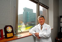
Stem Cells Heal Lungs of Newborn Animals

Study anticipated to benefit premature babies Saturday, 28 November 2009 Dr. Bernard Th̩baud lives in two very different worlds. As a specialist in the Stollery Children's Hospital's Neonatal Intensive Care Unit at the Royal Alexandra Hospital, he cares for tiny babies, many of whom struggle for breath after being born weeks before they are due. Across town, in his laboratory in the Faculty of Medicine & Dentistry at the University of Alberta, Dr. Th̩baud dons a lab coat and peers into a microscope to examine the precise effect of stem cells on the lungs. Today, with his scientific research being published in the American Journal of Respiratory and Critical Care Medicine, Dr. Th̩baud has made a significant leap to bridge the gap between those two worlds. An international team of scientists led by Dr. Th̩baud has demonstrated for the first time that stem cells protect and repair the lungs of newborn rats. "The really exciting thing that we discovered was that stem cells are like little factories, pumping out healing factors," says Dr. Th̩baud, an Alberta Heritage Foundation for Medical Research Clinical Scholar. "That healing liquid seems to boost the power of the healthy lung cells and helps them to repair the lungs." In this study, Th̩baud's team simulated the conditions of prematurity Рgiving the newborn rats oxygen. The scientists then took stem cells, derived from bone marrow, and injected them into the rats' airways. Two weeks later, the rats treated with stem cells were able to run twice as far, and had better survival rates. When Th̩baud's team looked at the lungs, they found the stem cells had repaired the lungs, and prevented further damage. "I want to congratulate Dr. Th̩baud and his team. This research offers real hope for a new treatment for babies with chronic lung disease," says Dr. Roberta Ballard, professor of paediatrics, University of California, San Francisco. "In a few short years, I anticipate we will be able to take these findings and begin clinical trials with premature babies." "The dilemma we face with these tiny babies is a serious one. When they are born too early, they simply cannot breathe on their own. To save the babies' lives, we put them on a ventilator and give them oxygen, leaving many of them with chronic lung disease," says Dr. Th̩baud. "Before the next decade is out I want to put a stop to this devastating disease." The research team includes physicians and scientists from Edmonton, Alberta, Tours, France, Chicago, Illinois, and Montreal, Quebec. The team is now investigating the long-term safety of using stem cells as a lung therapy. The scientists are examining rats at 3 months, and 6 months after treatment, studying the lungs, and checking their organs to rule out any risk of cancer. Dr. Th̩baud's team is also exploring whether human cord blood is a better option than bone marrow stem cells in treating lung disease. "We are also studying closely the healing liquid produced by the stem cells," says Dr. Th̩baud. "If that liquid can be used on its own to grow and repair the lungs, that might make the injection of stem cells unnecessary." Reference: Airway Delivery of Mesenchymal Stem Cells Prevents Arrested Alveolar Growth in Neonatal Lung Injury in Rats Timothy van Haaften, Roisin Byrne, Sebastien Bonnet, Gael Y. Rochefort, John Akabutu, Manaf Bouchentouf, Gloria J. Rey-Parra, Jacques Galipeau, Alois Haromy, Farah Eaton, Ming Chen, Kyoko Hashimoto, Doris Abley, Greg Korbutt, Stephen L. Archer, and Bernard Th̩baud Am. J. Respir. Crit. Care Med. 2009; 180: 1131-1142. ......... ZenMaster


For more on stem cells and cloning, go to CellNEWS at http://cellnews-blog.blogspot.com/ and http://www.geocities.com/giantfideli/index.html

Wednesday 25 November 2009

Research Teams Map Genetic, Genomic Patterns in Han Chinese Population

First 'genetic map' of Han Chinese may aid search for disease susceptibility genes Wednesday, 25 November 2009 A pair of papers in the American Journal of Human Genetics today is highlighting the genetic and genomic variation present within the Han Chinese population. It is the largest ethnic population in the world. In the first of these papers, a Genome Institute of Singapore-led team developed a genetic map of the Han Chinese population by genotyping thousands of individuals from across China. This first historical genetic variation map is providing insights into Han Chinese population structure and evolutionary history — for instance, revealing North-South population structure in China. And down the road, researchers say, the results should pave the way for genome-wide association and other studies in the population. Based on genome-wide DNA variation information in over 6,000 Han Chinese samples from 10 provinces in China, this new map provides information about the population structure and evolutionary history of this group of people that can help scientists to identify subtle differences in the genetic diversity of Asian populations. Understanding these differences may aid in the design and interpretation of studies to identify genes that confer susceptibility to such common diseases as diabetes in ethnic Chinese individuals. Understanding these differences also is crucial in exploring how genes and environment interact to cause diseases. With the genetic map, the GIS scientists were able to show that the northern inhabitants of China were genetically distinguishable from those in the south, a finding that seems very consistent with the Han Chinese's historical migration pattern. The genetic map also revealed that the genetic divergence was closely correlated with the geographic map of China. This finding suggests the persistence of local co-ancestry in the country. "The genome-wide genetic variation study is a powerful tool which may be used to infer a person's ancestral origin and to study population relationships," said Liu Jianjun, Ph.D., GIS Human Genetics Group Leader. "For example, an ethnic Chinese born and bred in Singapore can still be traced back to his or her ancestral roots in China," Dr. Liu said. "By investigating the genome-wide DNA variation, we can determine whether an anonymous person is a Chinese, what the ancestral origin of this person in China may be, and sometimes which dialect group of the Han Chinese this person may belong to.” "More importantly, our study provides information for a better design of genetic studies in the search for genes that confer susceptibility to various diseases," he added. Of particular interest to people in Singapore are the findings that while the majority of Singaporean Chinese hail from Southern China as expected, some have a more northern ancestral origin. GIS Executive Director Edison Liu, M.D., said: "Genome association studies have provided significant insights into the genes involved in common disorders such as diabetes, high cholesterol, allergies, and neurological disorders, but most of this work has been done on Caucasian populations.” "More recently, Dr. Liu Jianjun from our institute has been working with his Chinese colleagues to define the genetic causes of some of these diseases in Asian populations," the GIS Executive Director added. "This work refined those tools so that the results will not be obscured by subtle differences in the genetic diversity of Asian populations. In the process, Dr. Liu has reconstructed a genetic historical map of the Chinese people as they migrated from south to north over evolutionary time." "There are definite differences in genetic architecture between populations," noted Chia Kee Seng, M.D., Head, Department of Epidemiology & Public Health, National University of Singapore (NUS), and Director, NUS-GIS Centre for Molecular Epidemiology. "We have seen this in the Singapore Genome Variation Project, a Joint NUH-GIS effort. Understanding these differences is crucial in exploring how genes and environment interact to cause diseases," he added. The research results published in American Journal of Human Genetics is part of a larger ongoing project on the genome-wide association study of diseases among the Chinese population. The project is a collaboration between GIS and several institutions and universities in China. In Jan. 2009, Nature Genetics published the findings of researchers at the GIS and Anhui Medical University, China, on psoriasis, a common chronic skin disease. In that study, led by Dr. Liu Jianjun at the GIS and Dr. Zhang Xuejun at the Anhui Medical University, the scientists discovered a genetic variant that provides protection against the development of psoriasis. The collaboration's recent discovery of over a dozen genetic risk variants for systematic lupus erythematosus (SLE) in the Chinese population was published in Nature Genetics in Oct. 2009. In a second AJHG paper, a Chinese research team genotyped more than 1,700 Han Chinese individuals from dozens of sites in China as part of another study aimed at understanding the genetic and genomic patterns within the Han Chinese population. The researchers genotyped 1,721 Han Chinese samples at about 160,000 SNPs for this paper. They collected more than 1,500 of the samples, while 44 were collected through the Human Genome Diversity Panel project and 171 were collected in Beijing and Denver as part of the HapMap project. That team detected north-south stratification similar to that reported by the Singapore-led team, though they designated three main Han Chinese clusters from northern, southern, and central parts of China. Again, individuals from the cities — in this case Beijing, Shanghai, and Guangzhou — did not represent populations that were as homogenous as those in other locations were. The researchers also found some SNPs that were strongly differentiated in different parts of the country. For instance, they reported, the frequency of SNPs in the genes FADS2 and HCP5 varied from north to south. Based on several simulated GWAS, each involving 300 cases and 300 controls, the team suggested that even the relatively subtle genetic variation within China could lead to excess false-positive associations. "Although differences in allele frequencies among Han Chinese clusters are small, our study has demonstrated the importance of accounting for population stratification in order to reduce false-positive associations," the researchers wrote. Reference: Genetic Structure of the Han Chinese Population Revealed by Genome-wide SNP Variation Jieming Chen, Houfeng Zheng, Jin-Xin Bei, Liangdan Sun, Wei-hua Jia, Tao Li, Furen Zhang, Mark Seielstad, Yi-Xin Zeng, Xuejun Zhang, Jianjun Liu The American Journal of Human Genetics, 25 November 2009, doi:10.1016/j.ajhg.2009.10.016 Genomic Dissection of Population Substructure of Han Chinese and Its Implication in Association Studies Shuhua Xu, Xianyong Yin, Shilin Li, Wenfei Jin, Haiyi Lou, Ling Yang, Xiaohong Gong, Hongyan Wang, Yiping Shen, Xuedong Pan, Yungang He, Yajun Yang, Yi Wang, Wenqing Fu, Yu An, Jiucun Wang, Jingze Tan, Ji Qian, Xiaoli Chen, Xin Zhang, Yangfei Sun, Xuejun Zhang, Bailin Wu and Li Jin The American Journal of Human Genetics, 25 November 2009, doi:10.1016/j.ajhg.2009.10.015 ......... ZenMaster


For more on stem cells and cloning, go to CellNEWS at http://cellnews-blog.blogspot.com/

Wednesday 14 October 2009

New Strategy for Mending Broken Hearts?

Patch created to repair damaged heart tissue Wednesday, 14 October 2009 By mimicking the way embryonic stem cells develop into heart muscle in a lab, Duke University bioengineers believe they have taken an important first step toward growing a living "heart patch" to repair heart tissue damaged by disease. In a series of experiments using mouse embryonic stem cells, the bioengineers used a novel mould of their own design to fashion a three-dimensional "patch" made up of heart muscle cells, known as cardiomyocytes. The new tissue exhibited the two most important attributes of heart muscle cells – the ability to contract and to conduct electrical impulses. The mould looks much like a piece of Chex cereal in which researchers varied the shape and length of the pores to control the direction and orientation of the growing cells.

This is Brian Liau, left, and Nenad Buirsac. Credit: Duke Photography.The researchers grew the cells in an environment much like that found in natural tissues. They encapsulated the cells within a gel composed of the blood-clotting protein fibrin, which provided mechanical support to the cells, allowing them to form a three-dimensional structure. They also found that the cardiomyocytes flourished only in the presence of a class of "helper" cells known as cardiac fibroblasts, which comprise as much as 60 percent of all cells present in a human heart. "If you tried to grow cardiomyocytes alone, they develop into an unorganized ball of cells," said Brian Liau, graduate student in biomedical engineering at Duke's Pratt School of Engineering. Liau, who works in the laboratory of assistant professor Nenad Bursac, presented the results of his latest experiments during the annual scientific sessions of the Biomedical Engineering Society in Pittsburgh. "We found that adding cardiac fibroblasts to the growing cardiomyocytes created a nourishing environment that stimulated the cells to grow as if they were in a developing heart," Liau said. "When we tested the patch, we found that because the cells aligned themselves in the same direction, they were able to contract like native cells. They were also able to carry the electrical signals that make cardiomyocytes function in a coordinated fashion."
This is the mould used to create the heart patch. Credit: Brian Liau."The addition of fibroblasts in our experiments provided signals that we believe are present in a developing embryo," Liau said. The need for helper cells is not uncommon in mammalian development. For example, he explained, nerve cells need "sheathe" cells known as glia in order to develop and function properly. Bursac believes that the latest experiments represent a proof-of-principle advance, but said there are still many hurdles to overcome before such patches could be implanted into humans with heart disease. "While we were able to grow heart muscle cells that were able to contract with strength and carry electric impulses quickly, there are many other factors that need to be considered," Bursac said. "The use of fibrin as a structural material allowed us to grow thicker, three-dimensional patches, which would be essential for the delivery of therapeutic doses of cells. One of the major challenges then would be establishing a blood vessel supply to sustain the patch." This immunofluorescence staining image shows the cardiomyocytes in green and the fibroblasts interspersed around them in red. The cells are aligned around the central pore. Credit: Brian Liau.The researchers plan to test their model using non-embryonic stem cells. For use in humans, this is important for many reasons, both scientifically and ethically, Bursac said. Recent studies have demonstrated that some cells from human adults have the ability to be reprogrammed to become similar to embryonic stem cells. "Human cardiomyocytes tend to grow a lot slower than those of mice," Bursac said. "Since it takes nine months for the human heart to complete development, we need to find a way to get the cells to grow faster while maintaining the same essential properties of native cells." If they could use a patient's own cells, the patch would also evade an immune system reaction, Bursac added. .........
ZenMaster
For more on stem cells and cloning, go to CellNEWS at http://cellnews-blog.blogspot.com/ and http://www.geocities.com/giantfideli/index.html

Liver Cells Grown from Patients' Skin Cells

Treatment of liver diseases possible Wednesday, 14 October 2009 These are liver cells generated from skin that are shown to make human liver proteins Albumin in green and HNF4 in red. Credit: Medical College of Wisconsin.Scientists at The Medical College of Wisconsin in Milwaukee have successfully produced liver cells from patients' skin cells opening the possibility of treating a wide range of diseases that affect liver function. The study was led by Stephen A. Duncan, D. Phil., Marcus Professor in Human and Molecular Genetics, and professor of cell biology, neurobiology and anatomy, along with postdoctoral fellow Karim Si-Tayeb, Ph.D., and graduate student Ms. Fallon Noto. "This is a crucial step forward towards developing therapies that can potentially replace the need for scarce liver transplants, currently the only treatment for most advanced liver disease," says Dr. Duncan. Liver disease is the fourth leading cause of death among middle aged adults in the United States. Loss of liver function can be caused by several factors, including genetic mutations, infections with hepatitis viruses, by excessive alcohol consumption, or chronic use of some prescription drugs. When liver function goes awry it can result in a wide variety of disorders including diabetes and atherosclerosis and in many cases is fatal. The Medical College research team generated patient–specific liver cells by first repeating the work of James Thomson and colleagues at University of Wisconsin-Madison who showed that skin cells can be reprogrammed to become cells that resemble embryonic stem cells. They then tricked the skin–derived pluripotent stem cells into forming liver cells by mimicking the normal processes through which liver cells are made during embryonic development. Pluripotent stem cells are so named because of their capacity to develop into any one of eh more than 200 cell types in the human body. At the end of this process, the researchers found that they were able to very easily produce large numbers of relatively pure liver cells in laboratory culture dishes. "We were excited to discover that the liver cells produced from human skin cells were able to perform many of the activities associated with healthy adult liver function and that the cells could be injected into mouse livers where they integrated and were capable of making human liver proteins," says Dr. Duncan. Several studies have shown that liver cells generated from embryonic stem cells could potentially be used for therapy. However, the possible use of such cells is limited by ethical considerations associated with the generation of embryonic stem cells from preimplantation embryos and the fact that embryonic stem cells do not have the same genetic make-up as the patient. Although the investigations are still at an early stage the researchers believe that the reprogrammed skin cells could be used to investigate and potentially treat metabolic liver disease. The liver may be particularly suitable for stem-cell based therapies because it has a remarkable capacity to regenerate. It is interesting to note that the regenerative nature of the liver was referenced in the ancient Greek tale of Prometheus. When Prometheus was caught stealing the gift of fire from Zeus, he was punished by having his liver eaten daily by an eagle. This provided the eagle with an everlasting meal because each night the liver of Prometheus would re-grow. The liver is a central regulator of the body's metabolism and is responsible for controlling sugar and cholesterol levels, secretion of a variety of hormones, production of blood clotting factors, and has an essential role in preventing toxins from damaging other organs in the body. It is possible that in the future a small piece of skin from a patient with loss of liver function could be used to produce healthy liver cells, replacing the diseased liver with normal tissue. Recently, the National Institutes of Health's National Institute of Diabetes and Digestive and Kidney Diseases through the American Recovery and Reinvestment Act have provided the MCW researchers, in collaboration with Markus Grompe, M.D., at the Oregon Health and Science University, a $1 million research grant to pursue the possibility of using reprogrammed skin cells to study and treat metabolic liver disease. Using this support, as well as donations from individuals throughout Milwaukee, the Medial College researchers are currently producing reprogrammed cells from patients suffering from diabetes, hyperlipidemia, and hypercholesterolemia in an effort to identify new treatments for these diseases. ......... ZenMaster


For more on stem cells and cloning, go to CellNEWS at http://cellnews-blog.blogspot.com/ and http://www.geocities.com/giantfideli/index.html

Monday 12 October 2009

Jumping Genes, Gene Loss and Genome Dark Matter

New map of copy number variation in the human genome is a resource for human genetics Monday, 12 October 2009 In research published last week by Nature, an international team describes the finest map of changes to the structure of human genomes and a resource they have developed for researchers worldwide to look at the role of these changes in human disease. They also identify 75 'jumping genes' - regions of our genome that can be found in more than one location in some individuals. However, the team cautions that they have not found large numbers of candidates that might alter susceptibility to complex diseases such as diabetes or heart disease among the common structural variants. They suggest strategies for finding this 'dark matter' of genetic variation. Human genomes differ because of single-letter variations in the genetic code and also because whole segments of the code might be deleted or multiplied in different human genomes. These larger, structural differences are called copy number variants (CNVs). The new research to map and characterize CNVs is of a scale and a power unmatched to date, involving hundreds of human genomes, billions of data points and many thousands of CNVs. "This study is more than ten times as powerful as our first map, published three years ago," explains Dr Matt Hurles from the Wellcome Trust Sanger Institute and a leader on the project, "and much more detailed than any other. Importantly, we have also assigned the CNVs to a specific genetic background so that they can be readily examined in disease studies carried out by others, such as the Wellcome Trust Case Control Consortium.” "Nevertheless, we have not found large numbers of common CNVs that we can tie strongly to disease. There remains much to be discovered and much to understand and our freely available genotyped collection will drive that discovery." The results show that any two genomes differ by more than 1000 CNVs, or around 0.8% of a person's genome sequence. Most of these CNVs are deletions, with a minority being duplications.


Jumping genes.Chromosomes are shown colour-coded in the outermost circle. Inside are lines connecting the origin and the new location (where known) of 58 out of 75 putative inter-chromosomal duplications, coloured according to their chromosome of origin. Credit: Jan Aerts, Wellcome Trust Sanger Institute.


Two consequences are particularly striking in this study of apparently healthy people. First, 75 regions have jumped around in the genomes of these samples; second, more than 250 genes can lose one of the two copies in our genome without obvious consequences and a further 56 genes can fuse together potentially to form new composite genes. "This paper detailing common CNVs in different world populations, and providing the first glimpse into evolutionary biology of such class of human variation, is unquestionably one of the most important advances in human genome research since the completion of a reference human genome," says Professor James R. Lupski, Vice Chair of the department of Molecular and Human Genetics, Baylor College of Medicine, Houston, Texas. "It complements the cataloguing of single nucleotide variation delineated in the HapMap Project and will both enable some new approaches to, and further augment other studies of, basic human biology relevant to health and disease." "The genetic 'blueprint' of humans is the human genome," says Sir Mark Walport, Director of the Wellcome Trust. "But we are each unique as individuals, shaped by variation in both genome and environment. Understanding the variation amongst human genomes is key to understanding the inherited differences between each of us in health and disease. A whole new dimension has been added to our understanding of variation in the human genome by the identification of copy number variants." The results also give, for the first time, a minimum measure of the rate of CNV mutation: at least one in 17 children will have a new CNV. In many cases, that CNV will have no obvious clinical consequences. However, for some the effects are severe. In those cases the data are captured in the DECIPHER database, a repository of clinical information about CNVs designed to aid the diagnosis of rare disorders in young children. However, CNVs are not only about here and about now; they are also ancient legacies of how our ancestors adapted to their environments. Among the most impressive variations between populations are CNVs that modify the activity of the immune system, known to be evolving rapidly in human populations, and genes implicated in muscle function. The researchers propose that the consequences of these CNVs can be dissected in population studies. The team scanned 42 million locations on the genomes of 40 people, half of European ancestry and half of West-African ancestry. The scale of the method meant they could detect CNVs as small as 450 bases occurring in one in 20 individuals. However, the researchers concede that their map of common variants will not account for much of the 'dark matter' of the genome - the missing heritability where, despite diligent searches, genetic variants have not been found for common disease. "CNV studies have made huge advances in the past few years, but we are still looking only at the most common CNVs," explains Dr Steve Scherer of the Hospital for Sick Children, Toronto. "We suspect that there are many CNVs that have real clinical consequences that occur in perhaps one in 50 or one in 100 people - below the level we have detected.” "Success in the hunt for the missing genetic causes of common disease has become possible in the last few years and we expect to find more as higher resolution searches become possible." The research group have maximized the value of their research by not only mapping the CNVs, but by also genotyping them - assigning them to a specific genetic background that makes them readily useful in wider genetic studies, such as the Wellcome Trust Case Control Consortium. "We were determined to develop not only the map, but also to provide the resources that help other researchers and clinical cytogeneticists most rapidly use our CNV results," comments Dr Charles Lee, one of the project leaders from Brigham and Women's Hospital and Harvard Medical School in Boston, USA. "Already, the data that we have generated is benefiting other large-scale studies such as the 1000 Genomes Projects as well as making an enormous difference in the accurate interpretation of clinical genetic diagnoses.” "Nonetheless, the human CNV story is far from over." ......... ZenMaster


For more on stem cells and cloning, go to CellNEWS at http://cellnews-blog.blogspot.com/ and http://www.geocities.com/giantfideli/index.html